USG ABDOMEN- showed -
(1) Mild hepatomegaly with multiple rounded hypoechoic solid nodular masses ranging 15 to 45 mm size involving both lobes.
(2) A 6 cms long segment Jejunal loop hypoechoic concentric mural thickening ( 10 mm) in left upper abdomen with mild luminal dilatation ( classical aneurysmal dilatation with target sign ) . Rest bowel was normal.
(3) A large 4 cms sized rounded adjacent mesenteric nodal mass
(4) Slight omental thickening
(5) Mild ascites


Fig 2- Left upper abdominal US scan shows hypoechoic concentric Jejunal loop thickening with aneurysmal dilatation. Adjacent rounded hypoechoic mesenteric nodal mass also seen.
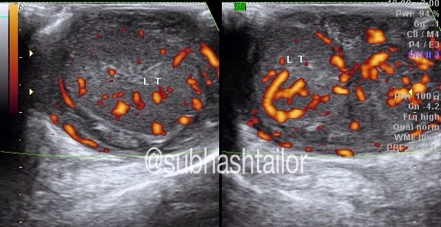
SCROTAL USG - findings were -
(1) Mildly bulky homogeneously hypoechoic both testes with mild hyperemia on doppler
(2) Diffusely hypoechoic epididymis
(3) Markedly thick inhomogenic hypoechoic & hyperemic extratesticular mass due to cord thickening, which is extending upto inguinal canal regions
(4) No hydrocele was present


PS -1) The case study is intended for medical professionals and imaging specialists for academic purposes
2) FNAC from hepatic nodule & testis showed NONHODGKIN'S LYMPHOMA
No comments:
Post a Comment