Sunday, November 10, 2013

EPIPLOIC APPENDAGITIS - An ultrasound diagnosis

Epiploic  appendages are peritoneal pouches that arise from the serosal surface of the colon. These are composed of adipose tissue and attached to colon by vascular  stalk  , multiple in count [ approx 100 ] ,& have a length range of  about 0.5–5 cm. Those located near the sigmoid colon are the largest in size . Epiploic appendages are usually not found near the rectum. The appendages are arranged in two rows: one row medial to the tenia libera, and the other lateral to the tenia omentalis of the colon .


Epiploic appendagitis is an uncommon self limiting inflammatory or  ischaemic condition affecting the colonic  epiploic appendage. Appendagitis  may be primary[ spontaneous ] or secondary to adjacent pathology , with more affection or predilection for sigmoid colon , presumably due to larger appendices. The condition most commonly manifests in the 4th to 5th decades of life, predominantly in men & obese individuals . Torsion of epiploic appendage pedicle , with resultant vascular occlusion or venous occlusion that leads to ischemia[ ischemic necrosis], has been implicated as the cause of acute epiploic appendagitis.  Acute epiploic appendagitis is also seen to be associated with  hernia, and unaccustomed exercise. Mostly  it is a self limiting  disease  , and  very uncommonly it may result in adhesion, bowel obstruction, intussusception, intraperitoneal loose body, peritonitis, and/or focal abscess formation.


Clinically patients present with acute abdominal pain and guarding in local area at the site of involvement [ that is usually lower quadrant , more common in left iliac fossa] . On the basis of clinical manifestations alone, acute epiploic appendagitis is mis-diagnosed in most of the cases , and or  often its being mistaken for acute diverticulitis.  Unlike acute epiploic appendagitis, acute diverticulitis is more likely to cause evenly distributed lower abdominal pain and  associated with nausea, fever, and leukocytosis.  On the contrary most patients with acute epiploic appendagitis do not manifest  any change in their bowel habits, and only a few cases report constipation or diarrhea.

Differential diagnosis ;
1] acute omental infarction
2] diverticulitis
3]sclerosing mesenteritis or mesenteric panniculitis
4] primary tumor or metastasis that involves peritoneum and mesocolon.
Ultrasound Findings
The site of maximum tenderness[mostly left iliac fossa] is the region of interest for ultrasound evaluation . There is evidence of a round to oval echogenic  noncompressible  mass noted , with no internal vascularity on doppler, and surrounded by a subtle hypoechoic line . The lesion is  typically 2 - 5 cm in maximal diameter adjacent and anterior to colon , and just deep to abdominal wall . The mass exert slight compressive effect on colonic loop  , and usually not  associated with colonic mural thickening  , or local or free peritoneal fluid .
In my series of 7 patients  [ 6 M & 2F, age range 35 to 48 yrs] , six cases presented with pain left iliac fossa , & one case with pain in right iliac fossa.  All cases were showed classical ultrasound findings of epiploic appendagitis with no bowel thickening or ascites . In all cases no leucocytosis was observed in lab. tests. In one case with pain right iliac fossa , the d/d was with acute appendicitis , but appendix and ileo-cecal region was seen separately & normally on ultrasound  .

Fig 1- Shows a normal  descending colonic epiploic appendage : An oval elongated echogenic structure [ arrow] attached to DC loop, nicely depicted due to presence of ascites[FL]


Fig 2 - Epiploic appendagitis, case A : LIF scan shows a well defined oval echogenic mass [bet cursors] surrounded by hypoechoic halo , just deep to abd wall 

Fig 3 - LIF scan of same case A , mass with no internal vascularity on power  doppler

Fig 4 - CT scan sagittal reconstruction image of  case A, shows a well defined oval hypodense fat density lesion along descending colonic loop[arrow within circle]

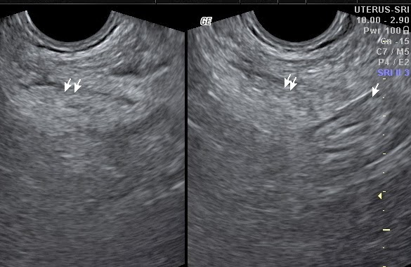
Fig 5 - Epiploic appendagitis, Case B, LIF scan shows a well defined  oval echogenic mass[ twin arrow] between sigmoid loop[single arrow] and abd wall

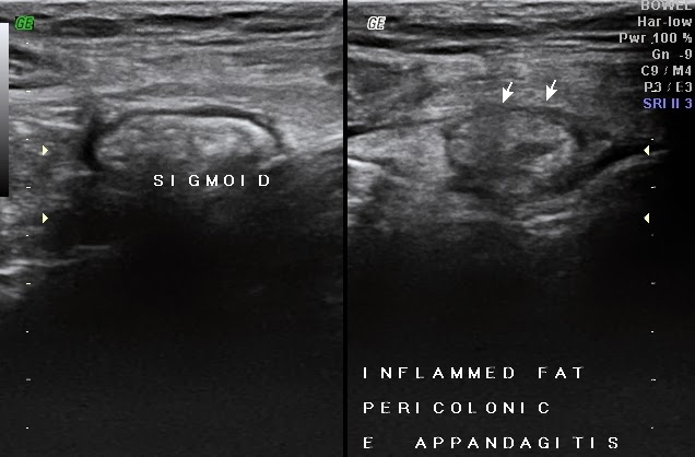
Fig 6 - Epiploic appendagitis, case C, LIF scan shows an oval echogenic mass with hypoechoic rim[arrows] adjacent to normally appearing sigmoid cooln


CT findings-
A fat-density ovoid lesion  adjacent to colon, size varying 1.5 - 3.5cm in diameter , with thin high-density rim , surrounding inflammatory fat stranding, and thickening of the adjacent peritoneum. A central hyperdense dot like area also appreciated representing the thrombosed vascular pedicle. As a chronic sequele an infarcted appendix epiploica  may calcify, and may detach to form an intraperitoneal loose body.

Treatment and prognosis

Epiploic appendagitis is a self limiting disease, and thus a correct diagnosis is important with imaging modalities to prevents unnecessary surgery. 
 Ref.

Tuesday, October 15, 2013

DUCT PAPILLOMA BREAST [ An ultrasound & color doppler diagnosis ]


Clinically duct pailloma causes nipple discharge  , which is usually blood stained in nature .

On ultrasound  often  a duct is seen dilated  with  a soft tissue  density intraductal mass [ echogenic mass or filling defect ] noted within it .  Some times  associated intraductal fluid with internal echos also present . Duct papilloma may be of varying size , but usually small  , solitary or multiple , and nonmobile mural adherent lesion with internal vascularity of Doppler [ suggests soft tissue nature ] . This  should be differentiated  from intraductal  infected slough debris or other nonviable matter . Duct papillomas are common in subareolar  & juxtaareolar areas , with some times dilated  duct is seen extending into nipple  . Biopsy is advised to detect any desmoplastic / neoplastic change.


Case – A 55 yrs old  female presented with H/O blood stained nipple discharge . Ultrasound scan showed  a dilated duct  extending from nipple to subareolar and adjacent area ,with a small soft tissue density / echogenic intraductal , mural adherent , nonmobile mass  seen as a filling defect [ Fig 1, 2 ] . The intraductal mass showed  internal vascularity on color and spectral Doppler  , suggesting viable soft tissue nature of the lesion  [ Fig 3 and 4 ]. 

Fig 1 - Nipple US scan shows elongated curved tubular ductal area [arrows]
Fig 2 - Subareolar US scan shows conti..dilated duct with a small echogenic intraductal mass[M], seen  as filling defect 
Fig 3-   The intraductal mass shows internal vascularity on power doppler
Fig 4 - Arterial waveform  signals depicted within vessel of intrductal mass[papilloma]


Monday, October 14, 2013

SONOGRAPHIC EVALUATION OF FRACTURE PENIS

ABSTRACT

This case report highlights various aspects of penile fracture, which is a rare urological emergency .  The incidence of penile fracture is slightly increasing these days, may be due to performance enhancing medicines.   It being a medical emergency patient must seek immediate medical support.  A swift diagnosis and management is essential to avoid undesirable after effects as discussed.  This presentation delineates on how sonography helps to make a complete diagnosis of this condition. 
Key words : Penile fracture , sonography , penile trauma, tunica albuginea

INTRODUCTION

Penile fracture is due to rupture of the tunica albuginea sheath covering the erectile corpora cavernosa.  It may involve one or both corpora cavernosa.  The condition is usually under reported due to ethical and phychological reasons observed by the patients .  The diagnosis of fracture penis is usually clinical .  The sonography is asked when the observations are  atypical , and to  confirm the diagnosis , and to evaluate site, size, orientation  and extend of tunical injury along with penile soft tissue status and hematoma.

CASE REPORT

A 36 years old man presented with development of  sudden painful swelling and deformity of the penis following sexual intercourse.  After having strong clinical suspicion of penile fracture, the urologist referred the case for sonographic evaluation .  Sonography of penis was done with  high resolution linear probe  in longitudinal , transverse, oblique and horizontal planes with the region of interest in detail.  USG showed diffuse skin and soft tissue thickening of penis with a small defect in the continuity of tunica albuginea [an echogenic line encircling hypoechoic cavernosa] of Left lateral corpora cavernosa of penile mid shaft [fig 1-3 ].  The defect was about 15 mm in size & transversely oriented  with a large inhomogenic hematoma noted overlying it.  The hematoma  was seen as inhomogenic echopoor & avascular mass adjacent to defect [ fig 1-3] .  Rest of the corpora and spongiosa appeared normal.  Both cavernosal arteries showed normal to little low resistance blood flow pattern [fig 4].   No evidence of corpora spongiosa or urethral injury was seen .  So, based on these ultrasound findings [ that is - cavernosal tunical sheath tear with associated hematoma ] a diagnosis of penile fracture was made  and a detailed report was furnished .  The case was then urgently operated by the urologist [ tunical repair and hematoma drainage ] and the findings were confirmed[fig 5-8 ].  Post operative recovery was uneventful , & the patient behaved normally for erectile function  in subsequent follow-up visits.
Fig 1 – Transverse US  penile scan shows defect in tunical sheath of left corpora cavernosa on lateral aspect [ arrow] with adjacent hematoma
Fig 2- Horizontally oriented longitudinal penile scan showing  tunical sheath defect [ arrow] & overlying penile shaft hematoma.
Fig 3- Both TS & LS color Doppler penile scans showing sheath defect , avascular hematoma  & average vascularity in rest of penile  shaft.
Fig 4 – Spectral Doppler display left cavernosal  artery
Fig 5 – Per op. photograph showing subcut. Penile shaft hematoma

Fig 6 – Per op. photograph depicting sheath defect

Fig 7 – Photograph after tunical sheath repair

Fig 8 – Photograph after complete  penile surgery

[ Operative photographs  courtesy – Dr.  Manish Bhansali ,M.S. , D.N.B.{Urology}, Bhilwara]


DISCUSSION

Penile shaft consist of two elongated columns of erectile tissue called corpora cavernosa placed dorsolaterally , and a corpus spongiosum ventrally  containing erectile tissue and urethra.  Each column is enclosed in tunica albuginea sheath which is extension of Buck’s fascia over these cylindrical structures [ 1, 2] .   During erection these corporas enlarges due to blood pooling in vascular sinusoids and the tunica albuginea is thinned .  Any extrinsic bending  force to erected penis causes rupture of tunica due to rapid rise of inside pressure in corporas.  Activities like masturbation , self manipulation, sexual intercourse , and rolling over bed, can cause penile trauma and result in fractures.  Vaginal intercourse being the most common cause [ 3 ] .  Increased use of drugs which enhance the duration of erection [ 4 ], pre-existing penile disease or periurethral infections [ 5 ] increase the chance of penile fractures.  The affected person usually complain [ 1 -2 , 5 ] hearing of , or feeling , a sudden crack  or snap in the penis with  sudden loss of erection  and resultant swollen, deviated, painful penis .  In most of the cases usually one side of distal two third of penile shaft is fractured with less than half of cavernosal circumference affected.  Associated hematoma may be intra- cavernosal , intra and or extra cavernosal or may extend to scrotum, suprabubic area, perineum, and in some cases , even upto thighs .  Associated urethral injury may present as hematuria and dysuria.
The diagnosis of penile fracture is usually clinical, however not  possible make or may be inconclusive sometimes , needs further imaging .  The modalities available are cavernosography, sonography [ USG ] and MRI to confirm and identify penile fractures.   MRI being multiplanar and  with good soft tissue resolution scores over other modalities , however , for practical purposes, sonography preferred over MRI as it is easily available , cost effective , portable radiation free , less time consuming and allows dynamic evaluation of the region of interest [ 6 ].  High resolution sonography clearly depicts penile soft tissue anatomy , outline of corporas including status of tunica albuginea sheath and associated hematoma . Loss of continuity or defect in tunica albuginea with associated hematoma signifies fracture of penile shaft [ as seen in this case] . 
These patients requires early surgical treatment in form of  hematoma removal and tunical repair , which has been stated better than conservative measures [ 3, 5 ] , as this reduces the chance of scarring , angulation deformity and painful erection syndrome consequences.  
This patient was immediately subjected to surgery, which was uneventful, & showed normal postoperative recovery. The patient gave history of  satisfactory penile erection in his follow up visits .
So, it is worth to know about this urogenital emergency by the Doctors , so as to make a swift diagnosis of “ fracture penis “ and to refer such a case for appropriate mamagement to avoid devastating physical, functional and psychological consequences.

CONCLUSION  

A case of penile fracture needs to be diagnosed and treated promptly to avoid complications .  Ultrasound is considered to be an imaging modality  of choice in emergency as it is noninvasive, cost effective , radiation free , easily and widely available and can be repeated even at bed side .   All Doctors must be aware  of this entity in all aspects including clinical perspective , knowing about imaging findings and realising urgent need for treatment to avoid complications.


REFERENCES

1. Bannister LH, Dyson M. Reproductive system. In: Williams PL, editor. Gray's anatomy. 38th ed. New York, NY: Churchill Livingstone; 1995. pp. 1847–1880.
2. Rosse C, Gaddum-Rosse P. Pelvis and perineum. In: Rosse C, Gaddum-Rosse P, editors. Hollinhead’s textbook of anatomy. 5th ed. Philadelphia, Pa: Lippincott-Raven; 1997. pp. 639–700.
3. Ruckle HC, Hadley HR, Lui PD. Fracture of penis: diagnosis and management. Urology.1992;40(1):33–35. [PubMed]
4. Blake SM, Bowley DMG, Dickinson A. Fractured penis: Another complication of SILDENAFIL.Grand Rounds. 2002;2:11–12.
5. Brotzman GL. Penile fracture. J Am Board Fam Pract. 1991;4(5):351–353. [PubMed]
6. Nomura JT, Sierzenski PR. Ultrasound diagnosis of penile fracture. J Emerg Med. 2010;38(3):362–365. [PubMed]

7. S. G. Kachewar et al , Ultrasound Evaluation Of Penile Fracture , Biomed Imaging & Interventional Journal  2011 oct-dec ; 7[4]: e27.

Conference Photographs


IRIA national conference 2011,New Delhi 

IRIA national conference 2012 Hyderabad 


RAJAPICON 2012 Bhilwara 

IRIA Northeast conference Guwahati Dec 2012

Felicitation at Guwahati conference 


IRIA national conference 2013 Indore


IRIA national conference Agra 2014

RAJRADICON JAIPUR 

RAJ RADICON 2014 Jodhpur

CME in Kota


IRIA national conference 2015 Kochi

IRIA national conference kochi - Memento by Dr Joe Antony


CME on Current trends in women s imaging- in GMCH Kota on 22march2015


IRIA state conference 12-13 September 2015 in Jaipur , speaking on HRSG Rotator Cuff 


IRIA national conference jan 2016  in Bhubneswar - Talk on " sonograsphy in pediatric GI anomalies " in Dr S K Sharma  US Symposium session


                                       @ Session IRIA National conferenca jan -2017 , Jaipur


@ Session IRIA National conferenca jan -2017 , Jaipur

                                      @ Session IRIA National conferenca jan -2017 , Jaipur

@ IRIA National conferenca jan -2017 , Jaipur

@ IRIA National conferenca jan -2017 , Jaipur


@ IRIA National conferenca jan -2017 , Jaipur


SONOFEST May 17 - Jodhpur

@ session SONOFEST May 17 - Jodhpur

@session SONOFEST May 17 - Jodhpur

@ session CME may 17 at RNT MC Udaipur

@ session CME may 17 at RNT MC Udaipur


 CME may 17 at RNT MC Udaipur


CME may 17 at RNT MC Udaipur