Sunday, December 7, 2014

COARCTATION OF AORTA DIAGNOSED BY RENAL DOPPLER

COARCTATION OF AORTA DIAGNOSED BY RENAL DOPPLER IN A HYPERTENSIVE CHILD

Coarctation of the aorta is an abnormal  narrowing of the aorta that occurs as a result of persistent muscular tissue in the region of the ligamentum arteriosus. Coarctation most commonly seen  opposite the ductus arteriosus, caudal to origin of the left subclavian artery and is called juxta-ductal coarctation. Coarctation is a cause of secondary hypertension, resulting in differential pressures in the upper and lower extremities.   

CASE DETAILS
A13 yr young boy with hypertension [ BP upper limb 150/98] , subjected to abdominal ultrasound with special study for renal Doppler . Abdominal organs were normal at USG. On renal 
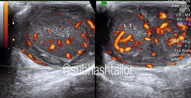
Doppler examination both sided main renal arteries were poorly seen & could not be clearly scanned. However in this case study  a low velocity low resistance  tardus -parvus waveform is seen in both sided intrarenal  renal arteries [fig 1 & 2 ].  The parvus tardus waveform of the renal artery is characterized by a slow rise of peak velocity distal to the stenosis, prolonged acceleration time and reduction of ipsilateral resistive index. This suggested that a stenosis proximal to the point being studied may be present. The supra-renal aorta showed a monophasic and dampened flow due to low flow velocities [ fig 3 ], further suggesting that a more proximal narrowing is likely , & that could be coarctation of aorta  . So , a possible diagnosis of coarctation of aorta was proposed .  Further examination by CT scan confirmed  the presence of juxta-ductal coarctation [ fig 4 & 5 ].

Fig 1-  Right intrarenal doppler shows tardus-parvus flow pattern . Note the decreased arterial peak systolic velocity & prolonged systolic peak acceleration time , which indicates proximal stenosis

Fig 2- Left intrarenal arterial doppler with similar tradus-parvus flow pattern

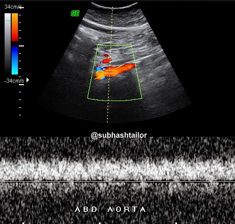
Fig 3 - Abdominal aortic doppler scan shows dampened monophasic blood flow spectrum due to low velocities , suggesting more proximal stenosis


Fig 4-  Sagittal CECT chest shows aortic stenosis [ c ] in juxta ductal isthmus of aorta [ AO ] caudal to left subclavian artery

Fig 5 - Coronal CECT chest shows juxta ductal aortic stenosis 


Take home point from this case: Look for a more proximal stenosis when abnormal bilateral renal artery waveforms [ tardus-parvus ] and abnormal aortic waveform [ dampened ] are noted. 


PS – The case study in intended for medical professionals & imaging specialists for academic purpose

Thursday, December 4, 2014

COMMON FEMORAL ARTERY THROMBOEMBOLISM

A 75 y female developed acute left lower limb pain with burning sensation . On examination the limb was cold with slight bluish discolouration of toes. Femoral pulse was feeble, & popliteal , posterior & anterior tibial , & dorsalis pedis pulses were not appreciable . Patient had attack of atrial fibrillation few days back . So , Clinically thromboembolism suspected of cardiac origin . Pt was hypertensive also .

AT Color Doppler - Left external iliac artery showed low velocity biphasic thump / short spectrum- s/o forward flow obstruction ( fig 1 ) . The left common femoral artery showed a Color & spectral signal void , with its lumen occluded by inhomogenically echogenic material s/o thrombosis ( fig 2&3 )  . The thrombus seen at bifurcation with partly extending into just proximal segments of SFA & DFA ( fig 4 ). Rest of arteries were collapsed with trace flow. 

The pt was then subjected to emergency surgical thromboembolectomy of femoral artery & recovered a large thromboembolus ( see fig -5&6)


Fig 1- Dual spectral doppler images with right EIA flow normal , & left EIA flow being short reduced spectrum s/o forward flow obstruction 

Fig2- Color Doppler scan of left common femoral artery showing intraluminal thrombosis ( Color void - arrows )

Fig3- Left common femoral artery shows spectral void 

Fig 4- Color Doppler scan shows thrombus extending into just proximal part of SFA & DFA


Fig 5- Sutured femoral artery after embolectomy. 

Fig 6- Post surgical embolectomy specimen recovered from femoral artery 

PS - 1) The case study is intended for medical professionals for academic purposes only.
 2) Thanks to Dr Anoop Gupta, cardiologist , at Sterling Hospital- Ahmedabad for feedback.

Thursday, November 27, 2014

GASTRIC TRICHOBEZOAR - an ultrasound diagnosis

GASTRIC TRICHOBEZOAR - An ultrasound diagnosis

BEZOAR- accumulation of undigested injested material within GIT. Gastric trichobezoar is formed by accumulation of injested hairs in stomach . It is hair ball mass usually take shape of stomach. It is seen commonly in mentally subnormal females who used to injest their own hairs.  Rapunzel syndrome is same condition in which a long tail like extension of bezoar noted reaching in small bowel causing its obstruction. Other types of bezoars are - phytobezoar ( formed by undigested food materials) ,  pharmacobezoar ( formed by drugs) , lactobezoar ( by milk products) .

CASE DETAILS
A  12 yr female child with pain & lumpish feel upper abdomen, occasional vomiting & loosing weight.
She was slightly mentally subnormal . On clinical examination upper abdominal lump was seen . Routine lab tests were noncontributory .
AT USG
A large complex mass was noted in upper abdomen , seen as a huge echodensity  in the region of stomach . The mass was filling & occupying entire stomach lumen with few visible thick gastric rugae . The mass was so large to cast a huge semilunar or crescentic echodensity with distal shadowing obscuring underlying details. No any other thing was seen except a huge echodense structure , that was traced in entire gastric cavity [ Fig 1-2] . Rest of the abdominal findings were normal on ultrasound . On the basis of sonographic findings a diagnosis of gastric trichobezoar was made , which was confirmed at surgery  [ Fig - ].

Fig 1- A midline sagittal US scan upper abomen shows part of left lobe liver , thick stomach rugae & a large semilunar echodensity [arrows ] filling entire stomach cavity

Fig 2 - Epigastric US scan shows a huge echodensity [ gastric bezoar ]  with distal shadowing

Fig 3- per operative images shows mass ( trichobezoar) distending  the stomach , & during its removal 

Fig 4- Post operative gross specimen (trichobezoar taken shape of stomach)


PS 1] The case study in intended for radiologists & sonologists for academic  purpose
     2 ] I expresss my sincere thanks to Dr Gaurav Bahety , M . Ch [pediatric surgery], Bhilwara -Rajasthan for operative details

Sunday, November 23, 2014

ULTRASOUND DIAGNOSIS OF SOLITARY SUBLINGUAL THYROID

A 10y boy diagnosed clinically & hormonally as hypothyroidism was evaluated with neck ultrasound. There was absence of normal thyroid in its infra laryngeal location . A well defined oval coarse hyperechoic spongiform nodular lesion noted in sublingual region midline supra hyoid location separate from both sublingual salivary glands . The lesion showed rim & internal vascularity on color doppler. No internal calcifications seen. In view of the nodule being in the track route of thyro-glossal duct, & absent normal thyroid in its location , a possible diagnosis of solitary sublingual thyroid was made ( Fig 1-4 ) . In vew of it's echo texture nodular spongiform appearance it might be adenomatous transforming . A thyroid isotope scan was suggested .


Fig 1- TS US neck scan shows absent normal thyroid in infra laryngeal location 

Fig 2- Coronal submental US scan shows a well defined spongiform nodule s/o sublingual thyroid tissue

Fig 3- The same nodule in midline sagittal scan seen in supra hyoid location 

Fig 4- color doppler shows rim & internal vasculature 

PS - The case study is intended for radiologists for academic purposes only 

Sunday, November 2, 2014

ULTRASOUND EVALUATION OF MUSCLE HERNIA

ULTRASOUND EVALUATION OF MUSCLE HERNIA


Most muscle hernias occur in the lower leg and affect the tibialis anterior muscle , and are attributed to occupational and sporting activities, trauma , chronic compartment syndrome, and weakness in the overlying fascia due to perforating vessels. It is seen  as a result of muscle protrusion through a defect in the muscle  fascia into the subcutaneous fat and eventually seen as  an overlying bulging soft-tissue mass , making slight contour hump. The adolescents or young adults are affected more. Clinically a swelling is  seen that usually enlarges when the affected muscle is contracted or the patient is in  standing or erect posture , and reduces when the muscle is relaxed .  Mostly muscle  hernias are asymptomatic and requires no treatment . Those with mild symptoms that may be relieved by support stockings. Patients with severe pain or cramps may require surgery.

At USG -  1 ] Focal thinning and slight elevation of the fascia.
                2] Focal fascial defect with protrusion or bulge  of muscle fibres through the defect  when the muscle                     is contracted , and making a contour hump. [ fig 1,2] 
               3] Prominent arterial pulsation on color or power Doppler - support the theory that muscle herniation                        occurs at sites of weakness in which vessels penetrate the fascia [ fig 3] 


                                          Fig 1 - A 20 yrs young boy presented with a small swelling at mid part of right anterolateral leg , which becomes conspicuous & painful on exercise or on continuous walking .  Long US scan shows focal bulge in tibialis anterior muscle [ arrows] on muscle contraction or on standing 

                                           
                                          Fig 2 - Both LS & TS ultrasound scans of the same case  , shows a small focal defect in the Fascia of tibialis  anterior muscle [ arrow in TS scan ]  with focal muscle fibres bulge or contour hump [ arrow in LS scan ]

                              
                                            Fig 3 - TS & LS ultrasound with color doppler scans shows a prominent vessel , likely perforating artery , at the site of fascial defect & muscle bulge or hernia  


Most muscle hernias do not require any  treatment [  or requires just reassurance] , but some painful cases may needs surgery. Importantly ultrasound is very helpful in apprehensive patients to exclude muscle tears and tumors . 


PS – This presentation is intended for academic purpose esp. for medical professionals & radiologists

Ref - American Journal of Roentgenology. 2003;180: 395-399

Thursday, October 30, 2014

DUCTAL CARCINOMA BREAST ENCASING THICK CALCIFIC WALLED TORTUROUS ARTERY- an ultrasound scan

A 50 yr female with lump right breast.
US scan showed a large well defined lobulated inhomogenic hypoechoic solid mass in central & outer region of right breast distorting glandular plane . The mass was encasing a prominently appreciable thick echogenic calcific walled tram track like tortuous structure within it.The structure had echogenic  tortuous branches which showed arterial colour & spectral signals on doppler & , this was a special  & unusual imaging feature in this case. No significant cystic or calcific foci noted within mass . The mass was abutting retro mammary muscle plane & extending deep to nipple . 
 
Fig - US scan right breast shows hypoechoic mass with echogenic elongated tortuous structure in it .

Fig 2- one another echogenic tram track like prominent branch also noted within mass which shows color flow signals on  color doppler , & this suggests vascular nature of this structure .


Fig 3- Spectral doppler interrogation shows arterial signals  

PS - 1) Biopsy proved the mass to be ductal carcinoma.
2) The case is presented to highlight this special imaging finding observed .
3) It is intended for radiologists for academic purpose only . 


Saturday, October 18, 2014

SEGMENTAL TESTICULAR INFARCT

Image Presentation

ACUTE SEGMENTAL TESTICULAR INFARCT -  mimic like a tumor on grayscale ultrasound , and color Doppler ultrasound  can clearly differentiate it from tumor .


Abstract
Segmental testicular infract is very uncommon pathology , & involves variable etiology , but commonly idiopathic . On grayscale ultrasound it appears as a focal inhomogenic mass which is difficult to be differentiated  from testicular tumor . However high resolution color Doppler ultrasound confidently diagnose it as an area of infarction & allows testis sparing surgery .

Introduction
Color Doppler ultrasound ( CDUS ) is very useful modality in evaluation of acute scrotum , and to  differentiate  between epididymoorchitis & torsion when symptoms may overlap  ( 2 ) .  The common conditions causing a painful scrotum  includes torsion & epididymoorchitis ,  and rarely tumor . Testicular tumor normally presents as a slow growing mass , rarely painful , and incidently discorved by the patient( 3 ) . On grayscale ultrasound tumor appears as an inhomogenic focal mass and CDUS demonstrates internal vascularity with malignant vascular pattern ( 4 ) . I present a case with painful testicular focal inhomogenic mass on grayscale ultrasound where CDUS allowed the diagnosis of segmental testicular infarction , rather than a testicular tumor , to be labelled .

Case report
A 46 years old man presented with a history of increasing right testicular pain for few days . The case was already  on antibiotics for presumed epididymoorchitis , however with no significant relief . On physical examination there was tenderness at the upper pole of right testis and epididymis , and a clinical diagnosis of epididymoorchitis was made.  In view of no relief in symptoms , the scrotal sonography was requested . Scrotal sonography was performed on GE – Logiq 400 pro  ultrasound system , using 8 to 11 MHZ. linear probe with small parts setting . The grayscale ultrasound examination revealed a focal enlarged inhomogenic area of  iso to hypoechoic echogenisity  at upper pole of right testis ( figure 1 ) . The CDUS failed to show any color flow signals within the mass but color flow signals were noted normal in rest of the testicular parenchyma ( figure 2 ) .  No any calcific focus or cystic change noted within lesion . No any other ultrasound evidence of epididymoorchitis was present . The possibility of an acute segmental testicular infract was made rather then a tumor because of absent color doppler signals in the focal abnormal area . The patient had normal complete blood count , and  was advised surgical exporation  by the referring doctor to exclude spermatic cord torsion as a cause for the abnormality . But  the patient  had refused for surgery , and  was  allowed to continue symptomatic medical treatment for some days .
After few weeks the referring doctor was contacted for follow up details of this case , and he ( referring doctor )  confirmed that the patient improved gradually and became symptom free with some residual testicular atrophy . The patient  was advised a review ultrasound  but he did not turn up .  
            
Discussion
Total testicular infarction is usually seen after torsion of spermatic cord , severe epididymoorchitis or trauma ( 2 ) . Segmental testicular infarction  is a rare entity and usually diagnosed by postorchidectomy histopathology  ( 5, 6 ) . The predisposing factors to segmental infarction  includes polycythemia ( 7 ) , intimal fibroplasia of spermatic artery ( 8 ) , sickle cell disease ( 9 ) , hypersensitivity angitits ( 10 ) and trauma , although most of the cases are idiopathic in origin ( 7 ) , as in this case . S. Sriprasad  et. al. reported a case  in BJR in 2001  with the same grayscale and CDUS  findings , which was proved to be a segmental testicular infarct histopathologically .  Scrotal sonography is valuable tool in differential diagnosis  of acute scrotum , & clearly differentiate  testicular torsion & infarct with high accuracy . In epididymoorchitis the testis and epididymis shows hyperemia , whereas absent or poor vascularity seen in torsion and infarct . The B- mode findings in acute testicular ischemia are enlarged and hypoechoic testis . CDUS helps to diagnose testicular torsion  where absent or poor blood flow noted in symptomatic testis  , and normal blood flow in contralateral  testis . The testicular tumors are usually seen as focal inhomogenic  or variable echotexture  masses with disordered / malignant internal vascularity on Doppler  ( 13 ) . On color Doppler , focal lesions larger than 16 mm , usually show raised and disordered blood flow ( 4 ) . Segmental testicular infarct also appears as focal mass of variable echogenicity with absent blood flow on Doppler ( 1 ) .  However cases with focal area of increased echogenicity  and poor or absent blood flow on CDUS were also reported and documented  in  segmental infarct ( 16 ) . This case showed  focal inhomogenic or variable echotexture mass at upper pole right testis with absent blood flow signals on CDUS , closely resembles the case reported by S. Sriprasad  et. al. . There was  focal enlargement of upper pole of testis , which may indicate acute nature of  the disease , because in chronic process the affected testis may appear small or shrunken ( 16 ) .  So with recent advances in probe technology and color Doppler sensitivity , it is  possible to document Intratesticular blood flow as well as vasculature pattern in a better way , which is  important particularly in differentiating a malignant mass from segmental infarction , as both appears identical on B – mode ultrasound , and thereby helpful in  suggesting a treatment  planning ( testis sparing & conservative management ) .


                   ( Figure 1 )    - Sagittal US image shows inhomogenic iso to hypoechoic mass like lesion 
                                         ( two small arrows ) involving upper pole of right testis    


               ( Figure 2 )  -  Color Doppler Us image of same testis shows absent color flow signals in the                                    abnormal area     s/o infarct , and normal blood flow signals in rest of the testis .

 PS ; The case study is based on imaging features & review literature , and intended for medical professionals & imaging specialists for academic purpose .


Referrences
1.      S .sriprasad  et al. Acute segmental testicular infarct : differentiation from tumour using high frequency colour Doppler ultrasound. BJR [74]2001,965-967
2.      Sidhu PS. Clinical and imaging features of testicular torsion: role of ultrasound. Clin Radiol 1999;54:343–52.[Medline]
3.      Morse MJ, Whitmore WF. Neoplasms of the testis. In: Walsh PC, Stamey TA, editors. Campbell's Urology (5th edn). Philadelphia, PA: WB Saunders Co., 1986:1535–82.
4.      Horstman WG, Melson GL, Middleton WD, Andriole GL. Testicular tumours: findings with color Doppler US. Radiology 1992;185:733–7.[Abstract/Free Full Text]
5.      Han DP, Dmochowski RR, Blasser MH, Auman JR. Segmental infarction of the testicle: atypical presentation of a testicular mass. J Urol 1994;151:159–60.[Medline]
6.      Costa M, Calleja R, Ball RY, Burgess N. Segmental testicular infarction. BJU Int 1999;83:525.[Medline]
7.      Jordan GH. Segmental hemorrhagic infarct of testicle. Urology 1987;29:60–3.[Medline]
8.      Brehmer-Andersson E, Andersson L, Johansson J. Hemorrhagic infarctions of testis due to intimal fibroplasia of spermatic artery. Urology 1985;25:379–82.[Medline]
9.      Holmes NM, Kane CJ. Testicular infarction associated with sickle cell disease. J Urol 1998;160:130.[Medline]
10.    Baer HM, Gerber WL, Kendall AR, Locke JL, Putong PB. Segmental infarct of the testis due to hypersensitivity angiitis. J Urol 1989;142:125–7.[Medline]
11.    Martin B, Conte J. Ultrasonography of the acute scrotum. J Clin Ultrasound 1987;15:37–44.[Medline]
12.    Middleton WD, Melson GL. Testicular ischemia: color Doppler sonographic findings in five patients. AJR 1989;152:1237–9.[Abstract/Free Full Text]
13.    Grantham JG, Charboneau JW, James EM, et al. Testicular neoplasms: 29 tumors studied by high-resolution US. Radiology 1985;157:775–80.[Abstract/Free Full Text]
14.    Gofrit ON, Rund D, Shapiro A, Pappo O, Landau EH, Pode D. Segmental testicular infarction due to sickle cell disease. J Ultrasound Med 1998;160:835–6.
15.    Flanagan JJ, Fowler RC. Testicular infarction mimicking tumour on scrotal ultrasound: a potential pitfall. Clin Radiol 1995;50:49–50.[Medline]
16.    Kramolowsky EV, Beauchamp RA, Milby WP. Color Doppler ultrasound for the diagnosis of segmental testicular infarction. J Urol 1993;150:972–3.[Medline]

17.    Bushby L, Sriprasad SI, Sidhu PS. Focal testicular abnormalities: evaluation of lesion vascularity using high frequency colour Doppler ultrasound. Eur J Ultrasound 2001;13:S30.

Sunday, October 12, 2014

POSSIBLE INTRAABDOMINAL TESTICULAR TUMOUR WITH PARAAORTIC LYMPHADENOPATHY - An ultrasound evaluation

POSSIBLE  INTRAABDOMINAL TESTICULAR TUMOR WITH  PARAAORTIC LYMPHADENOPATHY  - AN ULTRASOUND EVALUATION

CASE DETAILS -  A 34 years old man presented with lump lower abdomen &  mild pain ,  was referred for ultrasound abdomen  . No H/O vomiting , pyrexia , urinary or GI sign & symptoms was present  . Routine blood & urine tests were also non contributory .
AT USG - A large well defined  subtle inhomogenically hypoechoic  round to oval solid mass of about 95 x 54 x 65 mm size noted in suprapubic location abutting bladder wall. The mass was slightly mobile & extending towards right . No significantly appreciable internal vascularity was present . No internal calcifications or cystic changes were seen . This mass was assumed to be a neoplastic lesion of omento-mesenteric origion [ as it was slightly mobile ] or  nodal [ fig 1& 2] . There was also evidence of a well defined oval hypoechoic mass of about 29 x 14 mm size noted in upper paraaortic location at aorto-caval window , & was assumed to be lymphadenopathy [ fig 3] . Trace ascites was also present .  Rest of the abdominal organs were normal . In view of paraaortic lymphadenopathy the study was extended for scrotum to evaluate testes. Surprisingly both testes were absent in scrotum. Now retrospective H/O infertility was given by the  patient . A thorough search for undescended testes was made , and left testis of 31 x 16 x 21 mm size was found in intraabdominal location in left iliac fassa with homogenous echotexture [ fig -4 ] . Now the suprapubic mass was assumed to be  tumor development in right intraabdominal testis , as right testis was not separately seen . So on the basis of ultrasound findings a possible diagnosis of bilateral undesended intraabdominal testes with right testicular tumor and paraaortic lymphadenopathy was made.

Fig 1 - Suprapubic sag & transverse US scans showing a well defined oval solid mass abutting bladder wall
 [ possible right undescended intraabdominal testicular tumor]

Fig 2 - Color doppler scan of suprapubic mass , the mass did not revealed significantly appreciable color flow signals [ may be scanty poor flow ]

Fig 3 - Color doppler scan upper abdomen showing oval paraaortic lymphadenopathy in aorto-caval window
Fig 4 - Left iliac fossa US scan showing left intraabdominal testis

P S - The case study suggests possible diagnosis & is intended for medical professionals & imaging specialists for academic purpose

Tuesday, September 30, 2014

PELVIC ( BROAD LIGAMENT) HYDATID CYST- AN ULTRASOUND DIAGNOSIS

CASE DETAILS- A 20 yrs female with lower abdominal pain
AT US -  A large well defined rounded complex multicystic  mass noted in pelvis separate from normally appearing uterus and ovaries. The mass was harbouring multiple small rounded cysts ranging10 to 20 mm in size , mostly similar,  like bunch of grapes, with echofree interior . The lesion was seen superior & slightly right to uterus and bladder. No internal echos or vascularity noted within mass . The mass was showing slight double layered mural appearance at periphery. No solid components seen . No free fluid was noted in POD. No calcific foci or any other internal complexity was appreciated . No any other similar lesion was seen elsewhere in abdomen. On the basis of US findings a diagnosis of benign complex pelvic cyst , possibly Hydatid cyst of right broad ligament [ primary pelvic peritoneal hydatid disease ],  was made. Which was confirmed at surgery.

Fig 1- TVS scan showing a large complex multicystic pelvic mass with multiple rounded daughter cysts in it, & the mass was separate from uterus & both ovaries


Fig 2 - TVS scan showing double layered peripheral wall of the multicystic mass

Fig 3 - TVS scans showing normally & separately appearing uterus & both ovaries

AT SURGERY- Rt broad ligament Hydatid cyst with thin filmy adhesion with right ovary, bladder wall, adjacent bowel loops & abdominal wall noted . The cyst was successfully operated and removed.
Fig 4 - Photograh showing per operative view of pelvic cyst 

Fig 5-  Post op gross specimen showing ectocyst layer ( red piece) , endocyst layer( creamy white piece) , & multiple small daughter cysts in kidney tray .

PS- 1) The case study is intended for medical professionals & imaging specialists for academic interests.
2) Special thanks to Dr S P sharma ( Surgeon, Bhilwara (raj, India) for operative feedback.

Friday, September 26, 2014

MULTIORGAN ULTRASOUND FINDINGS IN A CHILD WITH NONHODGKIN'S LYMPHOMA


CASE DETAILS-  A 6 yrs old boy presented with wt loss, pain abdomen, occasional vomiting & painless scrotal swelling. 
USG ABDOMEN- showed -
(1) Mild  hepatomegaly with multiple rounded hypoechoic solid nodular masses ranging 15 to 45 mm size involving both lobes. 
(2) A 6 cms long segment Jejunal loop hypoechoic concentric mural thickening ( 10 mm) in left upper abdomen with mild luminal dilatation ( classical aneurysmal dilatation with target sign ) . Rest bowel was normal.
(3) A large 4 cms sized rounded adjacent mesenteric nodal mass
(4) Slight omental thickening
(5) Mild ascites

Fig 1- Hepatic US scan shows multiple rounded hypoechoic nodular metastatic masses


Fig 2-  Left upper abdominal US scan shows hypoechoic concentric Jejunal loop thickening with aneurysmal dilatation. Adjacent rounded hypoechoic mesenteric nodal mass also seen.

SCROTAL USG - findings were -

(1) Mildly bulky homogeneously hypoechoic both testes with mild hyperemia on doppler 
(2) Diffusely hypoechoic epididymis 
(3) Markedly thick inhomogenic hypoechoic & hyperemic extratesticular mass due to cord thickening, which is extending upto inguinal canal regions
(4) No hydrocele was present

Fig 3 - Scrotal US & color doppler scan showing homogeneously hypoechoic & hyperemic testis , & extratesticular masses due to cord & epididymal swelling.

Fig 4 - scrotal US scan showing hypoechoic testis & extratesticular masses due to cord and epididymal infiltration 

Fig 5- Quad US & color doppler images of both sided  supratesticular spermatic cords showing hypoechoic & hyperemic thickening of entire cord extending upto inguinal canal.

PS -1) The case study is intended for medical professionals and imaging specialists for academic purposes 
2) FNAC from hepatic nodule & testis showed NONHODGKIN'S LYMPHOMA 

Wednesday, September 10, 2014

ULTRASOUND DIAGNOSIS OF DUODENAL ULCER IN A PEDIATRIC CASE

CASE DETAILS : -
A 13 yrs old child with h/o chr pain upper abd , occasional vomiting , & had one episode of malena  , was referred for ultrasound . The case was evaluated with ultrasound with fasting status & allowed to sip some water to make upper GI window . The solid organs were normal . No small bowel dilatation was seen . The GEJ & stomach were also normal in mucosal pattern & appearance . The duodenal evaluation showed eccentric focal mural hypoechoic thickening ( 6 mm) in about15 to 20 mm circumferential area in its proximal part . There was a classical mucosal defect or focal depression noted in this region resembling an ulcer crater , harbouring specks of air foci in it ( fig 1). The lesion was constantly persistent on imaging  . Focal luminal & mural / mucosal distortion was also observed ( fig 1 ) . Multiple episodes of hyper duodeno-gastric regurgitation of fluid were also noted during real time scan . So on the basis of classical ultrasound findings a diagnosis of duodenal ulcer was made , which was subsequently confirmed on endoscopy ( fig 2 )
Fig 1- Epigastric midline slightly oblique US scan showing focal duodenal eccentric hypoechoic mural thickening encasing a classical depressive lesion s/o ulcer ( arrows)
Fig 2- Upper GI endoscopic images showing classical duodenal ulcer in frontal & profile views .
Fig 3- Collage showing US & Endoscopic images to highlight identical appearance of duodenal ulcer . 

PS - Endoscopic f/up : Dr S Bohra , Apollo Hospital , Ahmedabad .

Sunday, August 31, 2014

ULTRASOUND EVALUATION OF CA RECTUM

CASE HISTORY - A 40 yrs female with recurrent bleeding per rectum & lower abd pain .
AT USG -   On trans abd ultrasound focal iso to hypo & hyperechoic rounded lesions of variable size noted in liver with hypoechoic halo around ,s/o mets . Rest of abd scan was normal for abd viscera . To evaluate ano-colorectal region a non gyne TVS applied . Non gyne TVS application showed irregular hypoechoic mural thickening in rectum just above anorectal junction involving about 55 mm long segment with mural thickness ranging 10 to 14 mm distorting layer & mucosal pattern. The lesion was hyperemic on color Doppler. A tiny hypoechoic mural nodule also noted adjacently & separately   likely s/o satellite metastatic lesion . Multiple rounded hypoechoic perirectal lymphadenopathy also noted  . In view of above findings the diagnosis of Ca rectum was made with adjacent satellite mural lesion , perirectal metastatic lymphadenopathy  & hepatic metastasis .

Fig 1 - Hepatic scan showing mets

Fig 2- TVS scan with rectal evaluation showing perirectal nodes .

Fig 3- TVS scan showing irregular hypoechoic rectal wall thickening distorting gut signature  & adjacent tiny mural satellite lesion.


Fig 4- On color Doppler the rectal thickening showed hyperemia .
 
  This is how ultrasound completed the diagnosis & TVS helped in rectal evaluation in proper . 

PS - Histopathology revealed adeno ca rectum .

Sunday, August 24, 2014

ULTRASOUND IN NEONATAL HIRSCHSPRUNG DISEASE


CASE HISTORY 
A  20 days old neonate with distension of abdomen , at ultrasound showed  - slight narrowing of rectum ( dotted arrow )  & gross distension of sigmoid & descending colon ( arrow )  , & mild distension of entire colon. Rest of the abdomen was unremarkable  . On the basis of USG findings low (rectal) obstruction suspected with colonic distension , & possibility of Hirschsprung disease proposed .  On barium enema study classical findings of narrowing with coning ( zone of transition ) & distension of sigmoid noted , which supported further the diagnosis . The case underwent primary colostomy with rectal biopsy , which confirmed aganglionic nature Hirschsprung disease . 

Fig-1  suprapubic US scan shows rectal narrowing with coning ( dotted arrow ) , & distended sigmoid ( small arrow ) . UB seen empty . The image classically similar  to barium study .

Fig 2 - Quad US scan shows distended sigmoid , descending  & other parts of colon with rectal narrowing . 


Fig 3 - Lateral barium enema radiograph


Fig 4- AP barium enema radiograph 

Both fig 3 & 4 barium enema radiographs   
shows narrowing of rectum with coning / funnelling of zone of transition & distended sigmoid colon , classical for Hirschsprung  disease .  

Case 2-
Fig 5- Sono-Recto-sigmoidogram,showing rectal narrowing / stricture with sigmoid fullness-classically resembling with barium study consistent with possible diagnosis of "Hirschsprung disease " (story of a 30 days neonate with constipation & abdominal distension)

TAKE HOME- To diagnose Hirschsprung disease in neonatal period is quite feasible and rewarding. The liquid meconium in colon & recto-sigmoid region acts as a contrast ( echogenic liquid in bowel) during sonography & can delineate the rectal narrowing ( stricture as beak sign) with proximal sigmoid distension. In the late age the constipated & fecal matter loaded sigmoid obscures the stricture details due to fecal matter & gas shadowing .

PS - Barium enema radiographic images  & operative f/up courtesy : Dr Gaurav Bahety , M. Ch. pediatric surgeon , Bhilwara ( raj , India )