Tuesday, September 30, 2014

PELVIC ( BROAD LIGAMENT) HYDATID CYST- AN ULTRASOUND DIAGNOSIS

CASE DETAILS- A 20 yrs female with lower abdominal pain
AT US -  A large well defined rounded complex multicystic  mass noted in pelvis separate from normally appearing uterus and ovaries. The mass was harbouring multiple small rounded cysts ranging10 to 20 mm in size , mostly similar,  like bunch of grapes, with echofree interior . The lesion was seen superior & slightly right to uterus and bladder. No internal echos or vascularity noted within mass . The mass was showing slight double layered mural appearance at periphery. No solid components seen . No free fluid was noted in POD. No calcific foci or any other internal complexity was appreciated . No any other similar lesion was seen elsewhere in abdomen. On the basis of US findings a diagnosis of benign complex pelvic cyst , possibly Hydatid cyst of right broad ligament [ primary pelvic peritoneal hydatid disease ],  was made. Which was confirmed at surgery.

Fig 1- TVS scan showing a large complex multicystic pelvic mass with multiple rounded daughter cysts in it, & the mass was separate from uterus & both ovaries


Fig 2 - TVS scan showing double layered peripheral wall of the multicystic mass

Fig 3 - TVS scans showing normally & separately appearing uterus & both ovaries

AT SURGERY- Rt broad ligament Hydatid cyst with thin filmy adhesion with right ovary, bladder wall, adjacent bowel loops & abdominal wall noted . The cyst was successfully operated and removed.
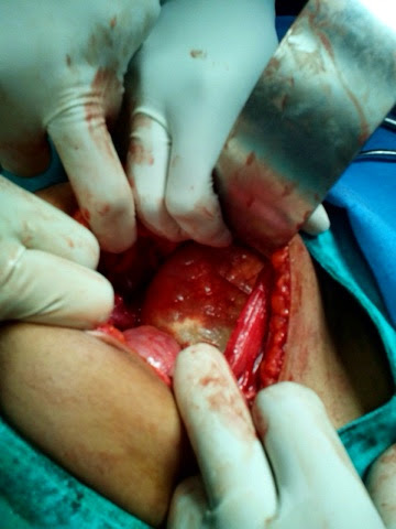
Fig 4 - Photograh showing per operative view of pelvic cyst 

Fig 5-  Post op gross specimen showing ectocyst layer ( red piece) , endocyst layer( creamy white piece) , & multiple small daughter cysts in kidney tray .
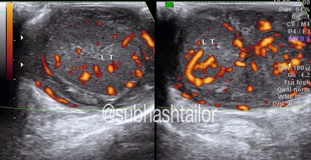
PS- 1) The case study is intended for medical professionals & imaging specialists for academic interests.
2) Special thanks to Dr S P sharma ( Surgeon, Bhilwara (raj, India) for operative feedback.

Friday, September 26, 2014

MULTIORGAN ULTRASOUND FINDINGS IN A CHILD WITH NONHODGKIN'S LYMPHOMA


CASE DETAILS-  A 6 yrs old boy presented with wt loss, pain abdomen, occasional vomiting & painless scrotal swelling. 
USG ABDOMEN- showed -
(1) Mild  hepatomegaly with multiple rounded hypoechoic solid nodular masses ranging 15 to 45 mm size involving both lobes. 
(2) A 6 cms long segment Jejunal loop hypoechoic concentric mural thickening ( 10 mm) in left upper abdomen with mild luminal dilatation ( classical aneurysmal dilatation with target sign ) . Rest bowel was normal.
(3) A large 4 cms sized rounded adjacent mesenteric nodal mass
(4) Slight omental thickening
(5) Mild ascites

Fig 1- Hepatic US scan shows multiple rounded hypoechoic nodular metastatic masses


Fig 2-  Left upper abdominal US scan shows hypoechoic concentric Jejunal loop thickening with aneurysmal dilatation. Adjacent rounded hypoechoic mesenteric nodal mass also seen.

SCROTAL USG - findings were -

(1) Mildly bulky homogeneously hypoechoic both testes with mild hyperemia on doppler 
(2) Diffusely hypoechoic epididymis 
(3) Markedly thick inhomogenic hypoechoic & hyperemic extratesticular mass due to cord thickening, which is extending upto inguinal canal regions
(4) No hydrocele was present

Fig 3 - Scrotal US & color doppler scan showing homogeneously hypoechoic & hyperemic testis , & extratesticular masses due to cord & epididymal swelling.

Fig 4 - scrotal US scan showing hypoechoic testis & extratesticular masses due to cord and epididymal infiltration 

Fig 5- Quad US & color doppler images of both sided  supratesticular spermatic cords showing hypoechoic & hyperemic thickening of entire cord extending upto inguinal canal.

PS -1) The case study is intended for medical professionals and imaging specialists for academic purposes 
2) FNAC from hepatic nodule & testis showed NONHODGKIN'S LYMPHOMA 

Wednesday, September 10, 2014

ULTRASOUND DIAGNOSIS OF DUODENAL ULCER IN A PEDIATRIC CASE

CASE DETAILS : -
A 13 yrs old child with h/o chr pain upper abd , occasional vomiting , & had one episode of malena  , was referred for ultrasound . The case was evaluated with ultrasound with fasting status & allowed to sip some water to make upper GI window . The solid organs were normal . No small bowel dilatation was seen . The GEJ & stomach were also normal in mucosal pattern & appearance . The duodenal evaluation showed eccentric focal mural hypoechoic thickening ( 6 mm) in about15 to 20 mm circumferential area in its proximal part . There was a classical mucosal defect or focal depression noted in this region resembling an ulcer crater , harbouring specks of air foci in it ( fig 1). The lesion was constantly persistent on imaging  . Focal luminal & mural / mucosal distortion was also observed ( fig 1 ) . Multiple episodes of hyper duodeno-gastric regurgitation of fluid were also noted during real time scan . So on the basis of classical ultrasound findings a diagnosis of duodenal ulcer was made , which was subsequently confirmed on endoscopy ( fig 2 )
Fig 1- Epigastric midline slightly oblique US scan showing focal duodenal eccentric hypoechoic mural thickening encasing a classical depressive lesion s/o ulcer ( arrows)
Fig 2- Upper GI endoscopic images showing classical duodenal ulcer in frontal & profile views .
Fig 3- Collage showing US & Endoscopic images to highlight identical appearance of duodenal ulcer . 

PS - Endoscopic f/up : Dr S Bohra , Apollo Hospital , Ahmedabad .