POSSIBLE INTRAABDOMINAL TESTICULAR TUMOR WITH PARAAORTIC LYMPHADENOPATHY - AN ULTRASOUND EVALUATION
CASE DETAILS - A 34 years old man presented with lump lower abdomen & mild pain , was referred for ultrasound abdomen . No H/O vomiting , pyrexia , urinary or GI sign & symptoms was present . Routine blood & urine tests were also non contributory .
AT USG - A large well defined subtle inhomogenically hypoechoic round to oval solid mass of about 95 x 54 x 65 mm size noted in suprapubic location abutting bladder wall. The mass was slightly mobile & extending towards right . No significantly appreciable internal vascularity was present . No internal calcifications or cystic changes were seen . This mass was assumed to be a neoplastic lesion of omento-mesenteric origion [ as it was slightly mobile ] or nodal [ fig 1& 2] . There was also evidence of a well defined oval hypoechoic mass of about 29 x 14 mm size noted in upper paraaortic location at aorto-caval window , & was assumed to be lymphadenopathy [ fig 3] . Trace ascites was also present . Rest of the abdominal organs were normal . In view of paraaortic lymphadenopathy the study was extended for scrotum to evaluate testes. Surprisingly both testes were absent in scrotum. Now retrospective H/O infertility was given by the patient . A thorough search for undescended testes was made , and left testis of 31 x 16 x 21 mm size was found in intraabdominal location in left iliac fassa with homogenous echotexture [ fig -4 ] . Now the suprapubic mass was assumed to be tumor development in right intraabdominal testis , as right testis was not separately seen . So on the basis of ultrasound findings a possible diagnosis of bilateral undesended intraabdominal testes with right testicular tumor and paraaortic lymphadenopathy was made.
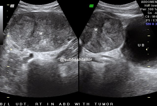 Fig 1 - Suprapubic sag & transverse US scans showing a well defined oval solid mass abutting bladder wall
[ possible right undescended intraabdominal testicular tumor]
 Fig 2 - Color doppler scan of suprapubic mass , the mass did not revealed significantly appreciable color flow signals [ may be scanty poor flow ]
 Fig 3 - Color doppler scan upper abdomen showing oval paraaortic lymphadenopathy in aorto-caval window
 Fig 4 - Left iliac fossa US scan showing left intraabdominal testis
P S - The case study suggests possible diagnosis & is intended for medical professionals & imaging specialists for academic purpose
CASE DETAILS - A 34 years old man presented with lump lower abdomen & mild pain , was referred for ultrasound abdomen . No H/O vomiting , pyrexia , urinary or GI sign & symptoms was present . Routine blood & urine tests were also non contributory .
AT USG - A large well defined subtle inhomogenically hypoechoic round to oval solid mass of about 95 x 54 x 65 mm size noted in suprapubic location abutting bladder wall. The mass was slightly mobile & extending towards right . No significantly appreciable internal vascularity was present . No internal calcifications or cystic changes were seen . This mass was assumed to be a neoplastic lesion of omento-mesenteric origion [ as it was slightly mobile ] or nodal [ fig 1& 2] . There was also evidence of a well defined oval hypoechoic mass of about 29 x 14 mm size noted in upper paraaortic location at aorto-caval window , & was assumed to be lymphadenopathy [ fig 3] . Trace ascites was also present . Rest of the abdominal organs were normal . In view of paraaortic lymphadenopathy the study was extended for scrotum to evaluate testes. Surprisingly both testes were absent in scrotum. Now retrospective H/O infertility was given by the patient . A thorough search for undescended testes was made , and left testis of 31 x 16 x 21 mm size was found in intraabdominal location in left iliac fassa with homogenous echotexture [ fig -4 ] . Now the suprapubic mass was assumed to be tumor development in right intraabdominal testis , as right testis was not separately seen . So on the basis of ultrasound findings a possible diagnosis of bilateral undesended intraabdominal testes with right testicular tumor and paraaortic lymphadenopathy was made.
[ possible right undescended intraabdominal testicular tumor]
P S - The case study suggests possible diagnosis & is intended for medical professionals & imaging specialists for academic purpose
