Showing posts with label Ultrasound. Show all posts

Sunday, April 19, 2015

ULTRASOUND IN HYDATID DISEASE


ULTRASOUND IN HYDATID DISEASE


Hydatid cyst is caused by  Echinococcus infection , resulting in cyst formation anywhere in the body. Humans are accidental host and the infection occurs by ingesting food contaminated with Echinococcus eggs.

Site preference -
  •  Liver – commonest
  •  Lung -  2nd commonest
  •  Spleen
  •  Central nervous system
  •  Retroperitoneal
  •  Musculoskeletal organs

Two main strains -

  1.  Echinococcus granulosus:  common
  2. Echinococcus alveolaris/multilocularis: less common but more invasive

The cysts usually have three components:
·   pericyst:  usually made up of inflammatory tissue of host origin
·   exocyst
·   endocyst: scolices (the larval stage of the parasite) and the laminated membrane  

The world health organization 2001 classification of hepatic hydatid cysts 

 CL - Unilocular anechoic cystic lesion without any internal echoes and septations
CE 1 -Uniformly anechoic cyst with fine echoes settled in it representing hydatid sand
CE 2 -Cyst with multiple septations giving it multivesicular appearance or rossette appearance
          or honey comb appearance with unilocular mother cyst . This stage is the active stage .
CE 3 - Unilocular cyst with daughter cysts with detached laminated membranes appearing as water
            lily sign , & this is the transitional stage of the cyst .
CE 4 - Mixed hypo and hyperechoic contents with absent daughter cysts, these contents give an
           appearance of ball of wool sign indicating the degenerative nature of the cyst .
CE 5 -Arch like thick partially or completely calcified wall , & this stage of cyst is inactive and
          infertile.

Morphological classification of cysts

Type I: simple cyst with no internal echos or  architecture
Type II: cyst with daughter cyst(s) + matrix echos
Type IIa: round daughter cysts at periphery
Type IIb: larger, irregularly shaped daughter cysts occupying entire volume of the mother                                   cyst
Type IIc: oval masses with scattered calcifications and occasional daughter cysts
Type III: calcified cyst (dead cyst)
Type IV: complicated cyst, e.g. ruptured cyst


IMAGE PRESENTATIONS

Case 1-Simple type 1 (morphological), CLtype (WHO), Active Hydatid Cyst

Fig 1-Hepatic sonogram shows a large well defined unilocular cystic mass in right lobe.The wall of the mass is smooth regular and slightly echogenic.This is a unilocular Simple Type 1  active Hydatid Cyst.

Fig 2- High Resolution hepatic sonogram reveals classical double echogenic layered appearance indicating inner endocyst and outer ectocyst (arrows).
  
 Fig 3- Axial CT scan image showes a large well defined unilocular rounded hypodense non enhancing mass in right lobe liver with fluid density.

 Fig 4-Coronal reconstruction CT image of the same case.

 Fig 5- Per operative photograph of the same case shows underlying cyst with bulging hepatic contour. 

 Fig 6-Per op photograph shows exposed interior of the cyst after incision and deflation.

 Fig 7-Shows removal of endocyst.

Fig 8- Gross Specimen of removed endocyst. 


Case 2- Type 2a (morphological), CE 2 (WHO), Active Cyst

Fig 9- Hepatic sonogram shows large hydatid cyst with multiple peripheral daughter cysts and central echogenic matrix- Spoke Wheel appearance


Case 3- Type 2b (morphological), CE 2 (WHO), Active Cyst

Fig 10- TVS pelvic scan shows large multiloculated adnexal mass with Honey Comb appearance.In this case mother cyst is full of multiple small daughter cysts. As both ovaries were seen separate and normal, the diagnosis of adnexal (likely broad ligament) pelvic hydatid cyst was made.

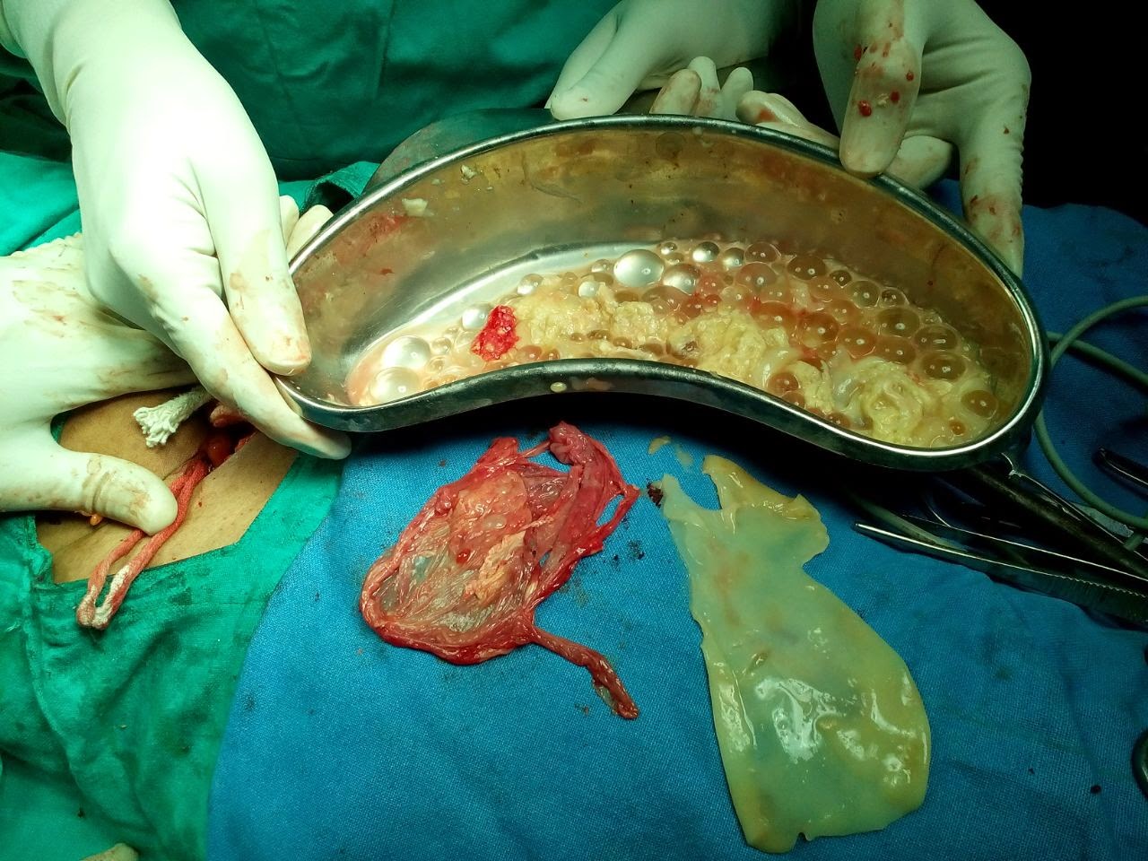
Fig 11- Post operative specimens of the above pelvic adnexal hydatid cyst case proved to be broad ligament hydatid cyst (one of the rare entity). Gross specimens showing ectocyst (red piece), endocyst (white piece) and kidney tray containing daughter cysts. 

Fig 12- Hepatic sonogram shows a large rounded complex cystic mass in left lobe with Honey Comb appearance.
Fig 13-Hepatic sonogram shows a large complex cystic mass with multiple small daughter cysts see as mobile sediments, likely suggests detached daughter cysts.

Case 4 -  CE 3 (WHO) type transitional stage hydatid cyst
.
Fig 14- Hepatic sonogram shows large complex cystic mass right lobe with detached endocyst layer appearing as echogenic curvilinear structure.

Fig 15- Hepatic sonogram of another case shows collapsed hydatid cyst with detached undulated endocyst.

Case 4- CE 4 type collapsed consolidating (solid appearing) inactive hydatid cyst.

Fig 16- Hepatic sonogram shows large rounded hyper echoic solid appearing mass with multiple hypoechoic curvilinear echos clustered within mass without daughter cysts. It is suggestive of consolidating inactive hydatid cyst.

Case 5 - Type 3 (morphological), CE 5 (WHO) type collapsed consolidating (solid appearing) inactive hydatid cyst.

Fig 17- Hepatic sonogram shows a well defined rounded mass with echogenic rim calcification casting distal shadowing. Here the lesion is inactive and infertile with calcifying nature.

Case 6 - Type 4 (morphological) Complicated hydatid cyst.

Fig 18- Hepatic Sonogram shows complex cystic mass left lobe liver full of internal echos and multiple bright foci with dirty down shadowing,suggestive of air. The case was earlier diagnosed as simple type hydatid cyst, which at present appears complicated by air and infection.


P.S.-This presentation is intended for medical professionals and imaging specialists for                       academic purposes. It needs discussion and further review of literature.
           - My special thanks to Dr.Gaurav Bahety M.Ch. (Ped. Surgeon) and Dr.S P Sharma                      (Gen.Surgeon) for operative feedback, and Dr.Kartikeya Nathiya, Bhilwara, for article                layout and crafting.


References
1] For more detail study – ref.  http://radiopaedia.org/articles/world-health-organization-2001-classification-of-hepatic-hydatid-cysts


Saturday, January 24, 2015

ULTRASOUND EVALUATION OF FOREARM ULNAR NERVE SCHWANNOMA

Schwannoma is an a well defined  encapsulated tumor of nerve sheath that grow eccentrically along the nerve axis, within the epineurium .  Schwannoma derive from cells representing the supporting tissue of a nerve, &  they typically appear as ovoid mass arising from the surface of a nerve separated from the unimpaired nerve fascicles . The nerve usually  stretched  & eccentrically courses over the capsule of the mass.  Sonography is usually unreliable in distinguishing between schwannomas and neurofibromas, as  both appears  as discrete homogeneous ovoid hypoechoic masses, with a healthy nerve at the proximal and distal ends of the mass .  A reliable sonographic diagnosis of a nerve tumor can only be made when the lesion found to be in continuity with nerve . The presence of cystic degeneration favours schwannoma rather than neurofibroma . Neurofibroma are surgically inseparable from the host nerve and can undergo malignant transformation ,where as schwannoa being discrete & eccentric ,  thus ,  often allowing the tumor to be surgically excised without loss of neurologic function.
CASE – A 35 y male presented with a small nodular swelling in distal part of  left forearm along ulnar aspest near wrist . The nodule was slightly painful and causes pain in ulnar half of hand on compressing it . No overlying skin changes or pulsations seen . No any pain noted on digital or wrist movements.
AT USG – A well defined encapsulated  oval hypoechoic solid nodular mass of about  16 x 10 x 12 mm size noted in inter mascular plane along ulnar vessels proximal to left wrist [ fig 1 ]. The lesion was  seen along ulnar nerve with neural fascicular continuity present &  established proximal & distal to the mass [ fig 2]. No significant internal vascularity or cystic or calcific foci noted . Adjacent ulnar vessels were  also seen normal . No muscle or bony or wrist  joint pathology seen . In view of encapsulated  focal nodular mass along ulnar nerve, a possible diagnosis of nerve tumor , likely schwannoma was made .  The lesion was proved ulnar nerve schwannoma  at surgery & biopsy [ fig 3 & 4 ].  

                                   Fig 1- LS & TS US scans of left distal forearm proximal to wrist,  showing a well defined hypoechoic  solid nodular mass adjacent to ulnar vessels [ along neurovascular bundle ] 

                                  Fig 2 - LS scan with conjoint image of the mass  , here the mass shows ulnar nerve fascicular continuity at its both proximal & distal ends , suggests the diagnosis of nerve tumor[ schwannoma ]

                                 Fig 3 - Per operative view of the nerve tumor . The tumor was removed without any neural damage .

                                     Fig 4 - Post operative gross specimen of dissected ulnar nerve sheath tumor [ schwannoma ]

PS – The case study in intended for medical professionals & imaging specialists for academic purpose.
  - My special thanks to Dr. Vishal Gupta , orthopaedic surgeon , Bhilwara [ raj., India ] , for operative feed back.


Read More: 1[  http://www.ajronline.org/doi/full/10.2214/ajr.182.1.1820123

                   2]  Peer et al J Ultrasound Med 21:315–322, 2002


Friday, January 16, 2015

NASOLABIAL CYST - An ultrasound evaluation

The nasolabial cyst is a rare non-odontogenic [ extraosseous ] cyst usually develops in the lower region of the nasal ala . Mostly its etiopathogenesis is uncertain .  It is more common in females at about age of forty . The cyst grows slowly and measures between 1.5 and 3 cm in size  It is characterized clinically by a floating structure in the nasolabial sulcus, which usually elevates the upper lip. It is also known as nasoalveolar cyst or Klestadt’s cyst . Its treatment is surgical excision through sublabial incision. Its recurrence is uncommon after complete removal .

CASE - This  is a 43 yrs female patient that presented with a firm swelling in the right alar region of the nose at nasolabial junction , slightly elevating the part of upper lip and nasal ala ; the clinical features suggested a nasolabial cyst.  HRSG scans revealed a well defined subtle thick and regular walled cystic lesion measuring  about 12 x 9 x 11 mm size in the  right ala of the nose [ fig 1-2 ].  No internal echos or septa or solid mural nodule is seen . It is slightly abutting underlying bone .

Fig 1- Slight oblique USG scan of right nasolabial junction showing  a well defined rounded cystic mass consistent with nasolabial cyst. No internal echos or septa or solid mural nodule is seen . Its wall is subtle thick & regular.


 Fig 2- Orthogonal US scans showing the dimensions of  the cyst

PS : The case is intended for academic purpose .


Ref : 1] http://radiopaedia.org/articles/nasolabial-cyst

Thursday, November 27, 2014

GASTRIC TRICHOBEZOAR - an ultrasound diagnosis

GASTRIC TRICHOBEZOAR - An ultrasound diagnosis

BEZOAR- accumulation of undigested injested material within GIT. Gastric trichobezoar is formed by accumulation of injested hairs in stomach . It is hair ball mass usually take shape of stomach. It is seen commonly in mentally subnormal females who used to injest their own hairs.  Rapunzel syndrome is same condition in which a long tail like extension of bezoar noted reaching in small bowel causing its obstruction. Other types of bezoars are - phytobezoar ( formed by undigested food materials) ,  pharmacobezoar ( formed by drugs) , lactobezoar ( by milk products) .

CASE DETAILS
A  12 yr female child with pain & lumpish feel upper abdomen, occasional vomiting & loosing weight.
She was slightly mentally subnormal . On clinical examination upper abdominal lump was seen . Routine lab tests were noncontributory .
AT USG
A large complex mass was noted in upper abdomen , seen as a huge echodensity  in the region of stomach . The mass was filling & occupying entire stomach lumen with few visible thick gastric rugae . The mass was so large to cast a huge semilunar or crescentic echodensity with distal shadowing obscuring underlying details. No any other thing was seen except a huge echodense structure , that was traced in entire gastric cavity [ Fig 1-2] . Rest of the abdominal findings were normal on ultrasound . On the basis of sonographic findings a diagnosis of gastric trichobezoar was made , which was confirmed at surgery  [ Fig - ].

Fig 1- A midline sagittal US scan upper abomen shows part of left lobe liver , thick stomach rugae & a large semilunar echodensity [arrows ] filling entire stomach cavity

Fig 2 - Epigastric US scan shows a huge echodensity [ gastric bezoar ]  with distal shadowing

Fig 3- per operative images shows mass ( trichobezoar) distending  the stomach , & during its removal 

Fig 4- Post operative gross specimen (trichobezoar taken shape of stomach)


PS 1] The case study in intended for radiologists & sonologists for academic  purpose
     2 ] I expresss my sincere thanks to Dr Gaurav Bahety , M . Ch [pediatric surgery], Bhilwara -Rajasthan for operative details

Saturday, October 18, 2014

SEGMENTAL TESTICULAR INFARCT

Image Presentation

ACUTE SEGMENTAL TESTICULAR INFARCT -  mimic like a tumor on grayscale ultrasound , and color Doppler ultrasound  can clearly differentiate it from tumor .


Abstract
Segmental testicular infract is very uncommon pathology , & involves variable etiology , but commonly idiopathic . On grayscale ultrasound it appears as a focal inhomogenic mass which is difficult to be differentiated  from testicular tumor . However high resolution color Doppler ultrasound confidently diagnose it as an area of infarction & allows testis sparing surgery .

Introduction
Color Doppler ultrasound ( CDUS ) is very useful modality in evaluation of acute scrotum , and to  differentiate  between epididymoorchitis & torsion when symptoms may overlap  ( 2 ) .  The common conditions causing a painful scrotum  includes torsion & epididymoorchitis ,  and rarely tumor . Testicular tumor normally presents as a slow growing mass , rarely painful , and incidently discorved by the patient( 3 ) . On grayscale ultrasound tumor appears as an inhomogenic focal mass and CDUS demonstrates internal vascularity with malignant vascular pattern ( 4 ) . I present a case with painful testicular focal inhomogenic mass on grayscale ultrasound where CDUS allowed the diagnosis of segmental testicular infarction , rather than a testicular tumor , to be labelled .

Case report
A 46 years old man presented with a history of increasing right testicular pain for few days . The case was already  on antibiotics for presumed epididymoorchitis , however with no significant relief . On physical examination there was tenderness at the upper pole of right testis and epididymis , and a clinical diagnosis of epididymoorchitis was made.  In view of no relief in symptoms , the scrotal sonography was requested . Scrotal sonography was performed on GE – Logiq 400 pro  ultrasound system , using 8 to 11 MHZ. linear probe with small parts setting . The grayscale ultrasound examination revealed a focal enlarged inhomogenic area of  iso to hypoechoic echogenisity  at upper pole of right testis ( figure 1 ) . The CDUS failed to show any color flow signals within the mass but color flow signals were noted normal in rest of the testicular parenchyma ( figure 2 ) .  No any calcific focus or cystic change noted within lesion . No any other ultrasound evidence of epididymoorchitis was present . The possibility of an acute segmental testicular infract was made rather then a tumor because of absent color doppler signals in the focal abnormal area . The patient had normal complete blood count , and  was advised surgical exporation  by the referring doctor to exclude spermatic cord torsion as a cause for the abnormality . But  the patient  had refused for surgery , and  was  allowed to continue symptomatic medical treatment for some days .
After few weeks the referring doctor was contacted for follow up details of this case , and he ( referring doctor )  confirmed that the patient improved gradually and became symptom free with some residual testicular atrophy . The patient  was advised a review ultrasound  but he did not turn up .  
            
Discussion
Total testicular infarction is usually seen after torsion of spermatic cord , severe epididymoorchitis or trauma ( 2 ) . Segmental testicular infarction  is a rare entity and usually diagnosed by postorchidectomy histopathology  ( 5, 6 ) . The predisposing factors to segmental infarction  includes polycythemia ( 7 ) , intimal fibroplasia of spermatic artery ( 8 ) , sickle cell disease ( 9 ) , hypersensitivity angitits ( 10 ) and trauma , although most of the cases are idiopathic in origin ( 7 ) , as in this case . S. Sriprasad  et. al. reported a case  in BJR in 2001  with the same grayscale and CDUS  findings , which was proved to be a segmental testicular infarct histopathologically .  Scrotal sonography is valuable tool in differential diagnosis  of acute scrotum , & clearly differentiate  testicular torsion & infarct with high accuracy . In epididymoorchitis the testis and epididymis shows hyperemia , whereas absent or poor vascularity seen in torsion and infarct . The B- mode findings in acute testicular ischemia are enlarged and hypoechoic testis . CDUS helps to diagnose testicular torsion  where absent or poor blood flow noted in symptomatic testis  , and normal blood flow in contralateral  testis . The testicular tumors are usually seen as focal inhomogenic  or variable echotexture  masses with disordered / malignant internal vascularity on Doppler  ( 13 ) . On color Doppler , focal lesions larger than 16 mm , usually show raised and disordered blood flow ( 4 ) . Segmental testicular infarct also appears as focal mass of variable echogenicity with absent blood flow on Doppler ( 1 ) .  However cases with focal area of increased echogenicity  and poor or absent blood flow on CDUS were also reported and documented  in  segmental infarct ( 16 ) . This case showed  focal inhomogenic or variable echotexture mass at upper pole right testis with absent blood flow signals on CDUS , closely resembles the case reported by S. Sriprasad  et. al. . There was  focal enlargement of upper pole of testis , which may indicate acute nature of  the disease , because in chronic process the affected testis may appear small or shrunken ( 16 ) .  So with recent advances in probe technology and color Doppler sensitivity , it is  possible to document Intratesticular blood flow as well as vasculature pattern in a better way , which is  important particularly in differentiating a malignant mass from segmental infarction , as both appears identical on B – mode ultrasound , and thereby helpful in  suggesting a treatment  planning ( testis sparing & conservative management ) .


                   ( Figure 1 )    - Sagittal US image shows inhomogenic iso to hypoechoic mass like lesion 
                                         ( two small arrows ) involving upper pole of right testis    


               ( Figure 2 )  -  Color Doppler Us image of same testis shows absent color flow signals in the                                    abnormal area     s/o infarct , and normal blood flow signals in rest of the testis .

 PS ; The case study is based on imaging features & review literature , and intended for medical professionals & imaging specialists for academic purpose .


Referrences
1.      S .sriprasad  et al. Acute segmental testicular infarct : differentiation from tumour using high frequency colour Doppler ultrasound. BJR [74]2001,965-967
2.      Sidhu PS. Clinical and imaging features of testicular torsion: role of ultrasound. Clin Radiol 1999;54:343–52.[Medline]
3.      Morse MJ, Whitmore WF. Neoplasms of the testis. In: Walsh PC, Stamey TA, editors. Campbell's Urology (5th edn). Philadelphia, PA: WB Saunders Co., 1986:1535–82.
4.      Horstman WG, Melson GL, Middleton WD, Andriole GL. Testicular tumours: findings with color Doppler US. Radiology 1992;185:733–7.[Abstract/Free Full Text]
5.      Han DP, Dmochowski RR, Blasser MH, Auman JR. Segmental infarction of the testicle: atypical presentation of a testicular mass. J Urol 1994;151:159–60.[Medline]
6.      Costa M, Calleja R, Ball RY, Burgess N. Segmental testicular infarction. BJU Int 1999;83:525.[Medline]
7.      Jordan GH. Segmental hemorrhagic infarct of testicle. Urology 1987;29:60–3.[Medline]
8.      Brehmer-Andersson E, Andersson L, Johansson J. Hemorrhagic infarctions of testis due to intimal fibroplasia of spermatic artery. Urology 1985;25:379–82.[Medline]
9.      Holmes NM, Kane CJ. Testicular infarction associated with sickle cell disease. J Urol 1998;160:130.[Medline]
10.    Baer HM, Gerber WL, Kendall AR, Locke JL, Putong PB. Segmental infarct of the testis due to hypersensitivity angiitis. J Urol 1989;142:125–7.[Medline]
11.    Martin B, Conte J. Ultrasonography of the acute scrotum. J Clin Ultrasound 1987;15:37–44.[Medline]
12.    Middleton WD, Melson GL. Testicular ischemia: color Doppler sonographic findings in five patients. AJR 1989;152:1237–9.[Abstract/Free Full Text]
13.    Grantham JG, Charboneau JW, James EM, et al. Testicular neoplasms: 29 tumors studied by high-resolution US. Radiology 1985;157:775–80.[Abstract/Free Full Text]
14.    Gofrit ON, Rund D, Shapiro A, Pappo O, Landau EH, Pode D. Segmental testicular infarction due to sickle cell disease. J Ultrasound Med 1998;160:835–6.
15.    Flanagan JJ, Fowler RC. Testicular infarction mimicking tumour on scrotal ultrasound: a potential pitfall. Clin Radiol 1995;50:49–50.[Medline]
16.    Kramolowsky EV, Beauchamp RA, Milby WP. Color Doppler ultrasound for the diagnosis of segmental testicular infarction. J Urol 1993;150:972–3.[Medline]

17.    Bushby L, Sriprasad SI, Sidhu PS. Focal testicular abnormalities: evaluation of lesion vascularity using high frequency colour Doppler ultrasound. Eur J Ultrasound 2001;13:S30.