Saturday, January 24, 2015

ULTRASOUND EVALUATION OF FOREARM ULNAR NERVE SCHWANNOMA

Schwannoma is an a well defined  encapsulated tumor of nerve sheath that grow eccentrically along the nerve axis, within the epineurium .  Schwannoma derive from cells representing the supporting tissue of a nerve, &  they typically appear as ovoid mass arising from the surface of a nerve separated from the unimpaired nerve fascicles . The nerve usually  stretched  & eccentrically courses over the capsule of the mass.  Sonography is usually unreliable in distinguishing between schwannomas and neurofibromas, as  both appears  as discrete homogeneous ovoid hypoechoic masses, with a healthy nerve at the proximal and distal ends of the mass .  A reliable sonographic diagnosis of a nerve tumor can only be made when the lesion found to be in continuity with nerve . The presence of cystic degeneration favours schwannoma rather than neurofibroma . Neurofibroma are surgically inseparable from the host nerve and can undergo malignant transformation ,where as schwannoa being discrete & eccentric ,  thus ,  often allowing the tumor to be surgically excised without loss of neurologic function.
CASE – A 35 y male presented with a small nodular swelling in distal part of  left forearm along ulnar aspest near wrist . The nodule was slightly painful and causes pain in ulnar half of hand on compressing it . No overlying skin changes or pulsations seen . No any pain noted on digital or wrist movements.
AT USG – A well defined encapsulated  oval hypoechoic solid nodular mass of about  16 x 10 x 12 mm size noted in inter mascular plane along ulnar vessels proximal to left wrist [ fig 1 ]. The lesion was  seen along ulnar nerve with neural fascicular continuity present &  established proximal & distal to the mass [ fig 2]. No significant internal vascularity or cystic or calcific foci noted . Adjacent ulnar vessels were  also seen normal . No muscle or bony or wrist  joint pathology seen . In view of encapsulated  focal nodular mass along ulnar nerve, a possible diagnosis of nerve tumor , likely schwannoma was made .  The lesion was proved ulnar nerve schwannoma  at surgery & biopsy [ fig 3 & 4 ].  

                                   Fig 1- LS & TS US scans of left distal forearm proximal to wrist,  showing a well defined hypoechoic  solid nodular mass adjacent to ulnar vessels [ along neurovascular bundle ] 

                                  Fig 2 - LS scan with conjoint image of the mass  , here the mass shows ulnar nerve fascicular continuity at its both proximal & distal ends , suggests the diagnosis of nerve tumor[ schwannoma ]

                                 Fig 3 - Per operative view of the nerve tumor . The tumor was removed without any neural damage .

                                     Fig 4 - Post operative gross specimen of dissected ulnar nerve sheath tumor [ schwannoma ]

PS – The case study in intended for medical professionals & imaging specialists for academic purpose.
  - My special thanks to Dr. Vishal Gupta , orthopaedic surgeon , Bhilwara [ raj., India ] , for operative feed back.


Read More: 1[  http://www.ajronline.org/doi/full/10.2214/ajr.182.1.1820123

                   2]  Peer et al J Ultrasound Med 21:315–322, 2002


Friday, January 16, 2015

NASOLABIAL CYST - An ultrasound evaluation

The nasolabial cyst is a rare non-odontogenic [ extraosseous ] cyst usually develops in the lower region of the nasal ala . Mostly its etiopathogenesis is uncertain .  It is more common in females at about age of forty . The cyst grows slowly and measures between 1.5 and 3 cm in size  It is characterized clinically by a floating structure in the nasolabial sulcus, which usually elevates the upper lip. It is also known as nasoalveolar cyst or Klestadt’s cyst . Its treatment is surgical excision through sublabial incision. Its recurrence is uncommon after complete removal .

CASE - This  is a 43 yrs female patient that presented with a firm swelling in the right alar region of the nose at nasolabial junction , slightly elevating the part of upper lip and nasal ala ; the clinical features suggested a nasolabial cyst.  HRSG scans revealed a well defined subtle thick and regular walled cystic lesion measuring  about 12 x 9 x 11 mm size in the  right ala of the nose [ fig 1-2 ].  No internal echos or septa or solid mural nodule is seen . It is slightly abutting underlying bone .

Fig 1- Slight oblique USG scan of right nasolabial junction showing  a well defined rounded cystic mass consistent with nasolabial cyst. No internal echos or septa or solid mural nodule is seen . Its wall is subtle thick & regular.


 Fig 2- Orthogonal US scans showing the dimensions of  the cyst

PS : The case is intended for academic purpose .


Ref : 1] http://radiopaedia.org/articles/nasolabial-cyst

Sunday, January 4, 2015

ULTRASOUND IN MID GUT MALROTATION

EPIGASTRIC WHIRLPOOL SIGN:
noted in midgut malrotation with transposition of SMA/SMV , & in some cases SMV winds around SMA in clockwise manner due to mesenteric twist & volvulus giving this whirlpool sign on ultrasound & color doppler  ( fig 1)
Fig 1- Epigastric  TS  US & Color doppler scan of a neonate with vomiting - shows classical whirlpool sign due midgut malposition & volvulus 

Fig 2- Midgut malrotation with volvulus at surgery ( photo courtesy- Dr Gaurav Bahety , pediatric surgeon, Bhilwara )

Sunday, December 7, 2014

COARCTATION OF AORTA DIAGNOSED BY RENAL DOPPLER

COARCTATION OF AORTA DIAGNOSED BY RENAL DOPPLER IN A HYPERTENSIVE CHILD

Coarctation of the aorta is an abnormal  narrowing of the aorta that occurs as a result of persistent muscular tissue in the region of the ligamentum arteriosus. Coarctation most commonly seen  opposite the ductus arteriosus, caudal to origin of the left subclavian artery and is called juxta-ductal coarctation. Coarctation is a cause of secondary hypertension, resulting in differential pressures in the upper and lower extremities.   

CASE DETAILS
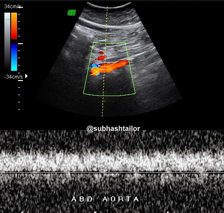
A13 yr young boy with hypertension [ BP upper limb 150/98] , subjected to abdominal ultrasound with special study for renal Doppler . Abdominal organs were normal at USG. On renal Doppler examination both sided main renal arteries were poorly seen & could not be clearly scanned. However in this case study  a low velocity low resistance  tardus -parvus waveform is seen in both sided intrarenal  renal arteries [fig 1 & 2 ].  The parvus tardus waveform of the renal artery is characterized by a slow rise of peak velocity distal to the stenosis, prolonged acceleration time and reduction of ipsilateral resistive index. This suggested that a stenosis proximal to the point being studied may be present. The supra-renal aorta showed a monophasic and dampened flow due to low flow velocities [ fig 3 ], further suggesting that a more proximal narrowing is likely , & that could be coarctation of aorta  . So , a possible diagnosis of coarctation of aorta was proposed .  Further examination by CT scan confirmed  the presence of juxta-ductal coarctation [ fig 4 & 5 ].

Fig 1-  Right intrarenal doppler shows tardus-parvus flow pattern . Note the decreased arterial peak systolic velocity & prolonged systolic peak acceleration time , which indicates proximal stenosis

Fig 2- Left intrarenal arterial doppler with similar tradus-parvus flow pattern

Fig 3 - Abdominal aortic doppler scan shows dampened monophasic blood flow spectrum due to low velocities , suggesting more proximal stenosis


Fig 4-  Sagittal CECT chest shows aortic stenosis [ c ] in juxta ductal isthmus of aorta [ AO ] caudal to left subclavian artery

Fig 5 - Coronal CECT chest shows juxta ductal aortic stenosis 


Take home point from this case: Look for a more proximal stenosis when abnormal bilateral renal artery waveforms [ tardus-parvus ] and abnormal aortic waveform [ dampened ] are noted. 


PS – The case study in intended for medical professionals & imaging specialists for academic purpose

Thursday, December 4, 2014

COMMON FEMORAL ARTERY THROMBOEMBOLISM

A 75 y female developed acute left lower limb pain with burning sensation . On examination the limb was cold with slight bluish discolouration of toes. Femoral pulse was feeble, & popliteal , posterior & anterior tibial , & dorsalis pedis pulses were not appreciable . Patient had attack of atrial fibrillation few days back . So , Clinically thromboembolism suspected of cardiac origin . Pt was hypertensive also .

AT Color Doppler - Left external iliac artery showed low velocity biphasic thump / short spectrum- s/o forward flow obstruction ( fig 1 ) . The left common femoral artery showed a Color & spectral signal void , with its lumen occluded by inhomogenically echogenic material s/o thrombosis ( fig 2&3 )  . The thrombus seen at bifurcation with partly extending into just proximal segments of SFA & DFA ( fig 4 ). Rest of arteries were collapsed with trace flow. 

The pt was then subjected to emergency surgical thromboembolectomy of femoral artery & recovered a large thromboembolus ( see fig -5&6)


Fig 1- Dual spectral doppler images with right EIA flow normal , & left EIA flow being short reduced spectrum s/o forward flow obstruction 

Fig2- Color Doppler scan of left common femoral artery showing intraluminal thrombosis ( Color void - arrows )

Fig3- Left common femoral artery shows spectral void 

Fig 4- Color Doppler scan shows thrombus extending into just proximal part of SFA & DFA


Fig 5- Sutured femoral artery after embolectomy. 

Fig 6- Post surgical embolectomy specimen recovered from femoral artery 

PS - 1) The case study is intended for medical professionals for academic purposes only.
 2) Thanks to Dr Anoop Gupta, cardiologist , at Sterling Hospital- Ahmedabad for feedback.

Thursday, November 27, 2014

GASTRIC TRICHOBEZOAR - an ultrasound diagnosis

GASTRIC TRICHOBEZOAR - An ultrasound diagnosis

BEZOAR- accumulation of undigested injested material within GIT. Gastric trichobezoar is formed by accumulation of injested hairs in stomach . It is hair ball mass usually take shape of stomach. It is seen commonly in mentally subnormal females who used to injest their own hairs.  Rapunzel syndrome is same condition in which a long tail like extension of bezoar noted reaching in small bowel causing its obstruction. Other types of bezoars are - phytobezoar ( formed by undigested food materials) ,  pharmacobezoar ( formed by drugs) , lactobezoar ( by milk products) .

CASE DETAILS
A  12 yr female child with pain & lumpish feel upper abdomen, occasional vomiting & loosing weight.
She was slightly mentally subnormal . On clinical examination upper abdominal lump was seen . Routine lab tests were noncontributory .
AT USG
A large complex mass was noted in upper abdomen , seen as a huge echodensity  in the region of stomach . The mass was filling & occupying entire stomach lumen with few visible thick gastric rugae . The mass was so large to cast a huge semilunar or crescentic echodensity with distal shadowing obscuring underlying details. No any other thing was seen except a huge echodense structure , that was traced in entire gastric cavity [ Fig 1-2] . Rest of the abdominal findings were normal on ultrasound . On the basis of sonographic findings a diagnosis of gastric trichobezoar was made , which was confirmed at surgery  [ Fig - ].

Fig 1- A midline sagittal US scan upper abomen shows part of left lobe liver , thick stomach rugae & a large semilunar echodensity [arrows ] filling entire stomach cavity

Fig 2 - Epigastric US scan shows a huge echodensity [ gastric bezoar ]  with distal shadowing

Fig 3- per operative images shows mass ( trichobezoar) distending  the stomach , & during its removal 

Fig 4- Post operative gross specimen (trichobezoar taken shape of stomach)


PS 1] The case study in intended for radiologists & sonologists for academic  purpose
     2 ] I expresss my sincere thanks to Dr Gaurav Bahety , M . Ch [pediatric surgery], Bhilwara -Rajasthan for operative details

Sunday, November 23, 2014

ULTRASOUND DIAGNOSIS OF SOLITARY SUBLINGUAL THYROID

A 10y boy diagnosed clinically & hormonally as hypothyroidism was evaluated with neck ultrasound. There was absence of normal thyroid in its infra laryngeal location . A well defined oval coarse hyperechoic spongiform nodular lesion noted in sublingual region midline supra hyoid location separate from both sublingual salivary glands . The lesion showed rim & internal vascularity on color doppler. No internal calcifications seen. In view of the nodule being in the track route of thyro-glossal duct, & absent normal thyroid in its location , a possible diagnosis of solitary sublingual thyroid was made ( Fig 1-4 ) . In vew of it's echo texture nodular spongiform appearance it might be adenomatous transforming . A thyroid isotope scan was suggested .


Fig 1- TS US neck scan shows absent normal thyroid in infra laryngeal location 

Fig 2- Coronal submental US scan shows a well defined spongiform nodule s/o sublingual thyroid tissue

Fig 3- The same nodule in midline sagittal scan seen in supra hyoid location 

Fig 4- color doppler shows rim & internal vasculature 

PS - The case study is intended for radiologists for academic purposes only