Sunday, July 5, 2020

Thursday, April 4, 2019
" Cervix sign" in congenital hypertrophic pyloric stenosis

SMA stenosis at origin on color & spectral Doppler


Sunday, June 24, 2018
APPENDICITIS WITH PERFORATION AT TIP
ULTRASOUND EVALUATION OF APPENDICITIS WITH PERFORATION AT TIP
Clinicals- F/70y with pain RIF

Fig1- High resolution ultrasound scan of RIF shows thick walled tubulo target like gut Lesion consistent with inflamed appendix . A small breach is seen in mural continuity at tip with perifocal small amount of fluid and inflamed fat ( arrows) - s/o perforation.

Saturday, June 23, 2018
MOREL LAVALLEE LESION ON ULTRASOUND
" MOREL LAVALLEE LESION"
Clinicals - H/O trauma
- Painful swelling anterior aspect of knee


MOREL LAVALLEE LESION - It is due to closed degloving soft tissue injury associated with trauma . In this lesion skin & subcutaneous fat abruptly separate from underlying deep fascia forming a hemolymphatic mass . Thigh at greater trochanteric area is the most common site .
Tuesday, January 9, 2018
ULTRASOUND CHEST- Empyema thoracis & Enlarged Thymus
ENCYSTED /LOCULATED INFECTED PLEURAL EFFUSION [ EMPYEMA THORACIS]

Fig 1- Utrasound image of left side chest showing thick walled loculated pleural effusion with internal echos and septas - indicative of empyema .

Fig 2- CXR PA of the same case showing homogeneous opacity left mid and lower zone with smooth upper margin . The opacity is obscuring cardiac and dome outline .
D/D - 1) Consolidation - point against - absent air bronchograms
2)- Pleural effusion - point against - no mass effect - this can be explained as it is loculated empyema
USG OF ENLARGED THYMUS

CXR: Shows upper mediastinal widening

USG scan(chest, left parmedian TS view) of the same pediatric case: Shows enlarged thymus as well defined soft tissue mass in the region of superior mediastinum retrosternal space.

USG scan(chest, left parmedian LS view) of the same pediatric case: Shows enlarged thymus as well defined soft tissue mass in the region of superior mediastinum retrosternal space, antero-superior to heart and great vessels.
Tuesday, July 25, 2017
TRANSPERINEAL US SCAN DEPICTING ANAL FISTULA WITH PERIANAL ABSCESS
CASE 1-
CLINICAL PROFILE - A young female c/o pain during defecation & some pus discharge per rectum
EXAMINATION - High resolution transperineal USG

CASE 2

Tuesday, May 10, 2016
SOFT TISSUE ULTRASOUND
MYOCYSTICERCOSIS
CASE 1 - A case with small tender swelling right pectoral region
 Fig 1- High resolution US scan of right pectoral region shows a well defined rounded cystic lesion within pectoralis major muscle with eccentric echogenic nidus - consistent with myocysticercosisi [ vesicular stage ] with subtle hypoechoic halo/edema around .
CASE 2
A young female presented with small slightly painful nodule left breast


CASE 1 - A case with small tender swelling right pectoral region

CASE 2
A young female presented with small slightly painful nodule left breast

Fig 1 - High resolution US image of left breast showing a well defined round to oval slightly thick walled cystic lesion with eccentric echogenic mural nodule , the findings are consistent with cysticercosis

Fig 2- High resolution US & Elastography image of the breast cysticercus cyst , with classical BGR pattern seen on elastography
Saturday, May 7, 2016
ULTRASOUND IN RIGHT ILIAC FOSSA PATHOLOGIES
ULTRASOUND IN POST APPENDECTOMY FECAL FISTULA
CASE - A case with RIF discharging lesion . H/O - appendectomy a month back .

Fig 1 - High resolution right iliac fossa US scan - shows a well defined hypoechoic track like lesion with central echogenicitis [ arrows ] indicative of air foci extending from cecal area to open externally through abdominal wall, s/o fecal fistula. The lumen of lesion is connected with lumen of cecum.
Friday, May 6, 2016
TESTICULAR DOPPLER ; VARICOCELES - COLOR & SPECTRAL DOPPLER

Fig 1- Color doppler scrotal scan showing dilated & tortuous venous vessels [ structures with red & blue color filled signals]

Saturday, April 30, 2016
OCULAR USG

Fig 1 - Ocular cysticercosis ; High resolution ocular USG scan depicting a well defined ring lesion posteromedial aspect of eye ball ,consistent with cysticercus cyst [ vesicular stage ] with eccentric echogenic nidus[scolex] . Retinal detachment is also evident.
[ Image courtesy: Dr Ajay Khandelwal , Alwar -Raj, India ]
DUODENAL USG
Duodenal ulcer disease
CASE 1

CASE 2
A young man with h/o chronic upper abdominal pain & occassional vomiting

also noted .

CASE 3
CLINICALS - A young lady c/o pain upper abdomen since 15 to 20 days , with occ vomiting .

Fig 1- Epigastric USscan depicting duodenum in transversa plane with echogenic mural lesion consistent with ulcer crater [ arrow]
Thursday, July 16, 2015
UTERINE AVMS WITH MYOMETRIAL PSEUDOANEURYSM
A young female with early pregnancy had h/o D&C done . She then developed excessive bleeding PV for 1 month.
This case was evacuated with TVS ....showed some retained products of conception, & multiple vascular cystic areas in juxtaposed myometrium with mosaic Color flow signals on Doppler. A large rounded unilocular intramural cystic mass was also noted anteriorly which showed color signals , s/o- pseudoaneurysm ( fig 1 to 4 ) .

Fig 1- TVS sag scan shows some retained products in endometrial cavity & a large round ed cystic mass in anterior uterine wall with mobile low level int. echoes .


Post gestational uterine AVMs are encountered in gynecological practice, but associated myometrial pseudoaneurysm is a rare entity .
PS : 1) CASE & images courtesy- Dr Renu Sharma - Radiologist ( Sikar, Rajasthan , India)
2) The case study is intended for imaging specialists & medical practitioners for academic purpose only.
3) It needs further discussion & review of literature
Wednesday, July 15, 2015
OCULAR LENS LUXATION
POSTERIOR DISLOCATION OF OCULAR LENS
CASE -
A 70y male sustained blunt ocular trauma with resulting loss of vision. HRSG of eye ball showed complete posterior dislocation of bulky thick walled ( cataractous ) lens [ fig 1& 2].
 Fig 1 - Ocular HRSG shows a biconvex echogenic structure lodged posteriorly in vitreous chamber near retina - dislocated bulky cataractous lens - likely due to rupture of weakened suspensory ligaments torned as a result of blunt ocular trauma
 Fig 2 - Ocular HRSG shows complete posterior luxation of lens
CASE -
A 70y male sustained blunt ocular trauma with resulting loss of vision. HRSG of eye ball showed complete posterior dislocation of bulky thick walled ( cataractous ) lens [ fig 1& 2].


Sunday, April 19, 2015
ULTRASOUND IN HYDATID DISEASE
ULTRASOUND IN HYDATID DISEASE
Hydatid cyst is caused by Echinococcus infection , resulting in cyst
formation anywhere in the body. Humans are
accidental host and the infection occurs by ingesting food contaminated with
Echinococcus eggs.
Site preference -
- Liver – commonest
- Lung - 2nd commonest
- Spleen
- Central nervous system
- Retroperitoneal
- Musculoskeletal organs
Two main strains -
- Echinococcus granulosus: common
- Echinococcus alveolaris/multilocularis: less common but more invasive
The cysts usually have three components:
· pericyst: usually made up of inflammatory
tissue of host origin
· exocyst
· endocyst: scolices (the larval stage of the parasite) and the
laminated membrane
The world
health organization 2001 classification of hepatic hydatid cysts
CL - Unilocular anechoic cystic lesion without
any internal echoes and septations
CE 1 -Uniformly anechoic cyst with
fine echoes settled in it representing hydatid sand
CE 2 -Cyst with multiple septations
giving it multivesicular appearance or rossette appearance
or honey comb appearance with unilocular mother cyst
. This stage is the active stage .
CE 3 - Unilocular cyst with daughter cysts with
detached laminated membranes appearing as water
lily sign , & this is the transitional
stage of the cyst .
CE 4 - Mixed hypo and hyperechoic contents with
absent daughter cysts, these contents give an
appearance of ball of wool sign indicating the
degenerative nature of the cyst .
CE 5 -Arch like thick partially or completely
calcified wall , & this stage of cyst is inactive and
infertile.
Morphological classification of cysts
Type I: simple cyst with no
internal echos or architecture
Type II: cyst with daughter
cyst(s) + matrix echos
Type IIa: round daughter cysts at periphery
Type IIb: larger, irregularly shaped daughter
cysts occupying entire volume of the mother cyst
Type IIc: oval masses with scattered calcifications and
occasional daughter cysts
Type III: calcified cyst
(dead cyst)
Type IV: complicated cyst, e.g. ruptured cyst
Type IV: complicated cyst, e.g. ruptured cyst
IMAGE PRESENTATIONS
Case 1-Simple type 1 (morphological), CLtype (WHO), Active Hydatid Cyst

Fig 1-Hepatic sonogram shows a large well defined unilocular cystic mass in right lobe.The wall of the mass is smooth regular and slightly echogenic.This is a unilocular Simple Type 1 active Hydatid Cyst.

Fig 2- High Resolution hepatic sonogram reveals classical double echogenic layered appearance indicating inner endocyst and outer ectocyst (arrows).

Fig 3- Axial CT scan image showes a large well defined unilocular rounded hypodense non enhancing mass in right lobe liver with fluid density.

Fig 4-Coronal reconstruction CT image of the same case.

Fig 5- Per operative photograph of the same case shows underlying cyst with bulging hepatic contour.

Fig 6-Per op photograph shows exposed interior of the cyst after incision and deflation.

Fig 7-Shows removal of endocyst.

Fig 8- Gross Specimen of removed endocyst.
Case 2- Type 2a (morphological), CE 2 (WHO), Active Cyst

Fig 9- Hepatic sonogram shows large hydatid cyst with multiple peripheral daughter cysts and central echogenic matrix- Spoke Wheel appearance
Case 3- Type 2b (morphological), CE 2 (WHO), Active Cyst

Fig 10- TVS pelvic scan shows large multiloculated adnexal mass with Honey Comb appearance.In this case mother cyst is full of multiple small daughter cysts. As both ovaries were seen separate and normal, the diagnosis of adnexal (likely broad ligament) pelvic hydatid cyst was made.
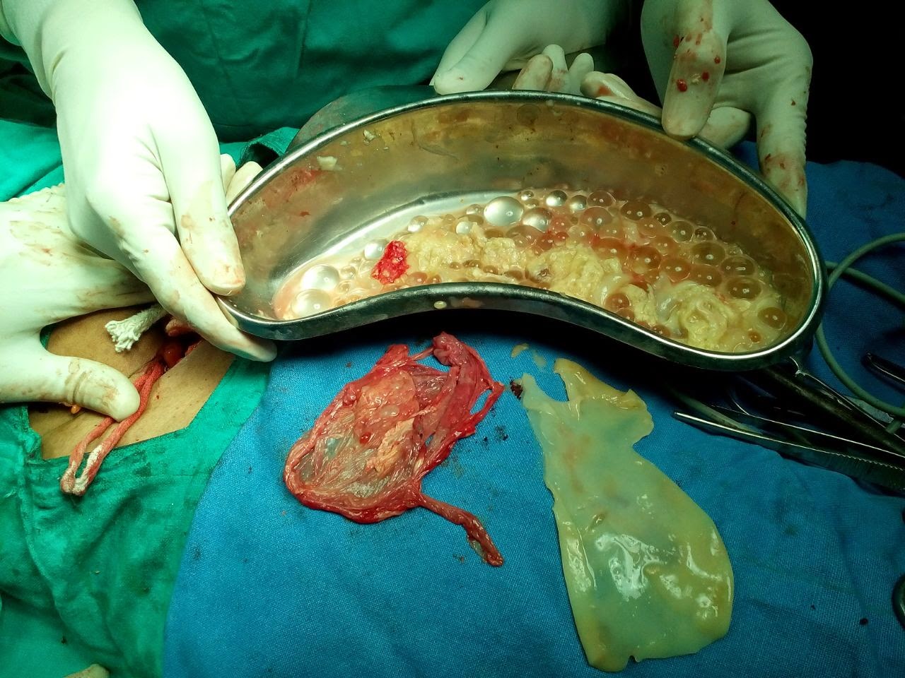
Fig 11- Post operative specimens of the above pelvic adnexal hydatid cyst case proved to be broad ligament hydatid cyst (one of the rare entity). Gross specimens showing ectocyst (red piece), endocyst (white piece) and kidney tray containing daughter cysts.
.jpg)
Fig 12- Hepatic sonogram shows a large rounded complex cystic mass in left lobe with Honey Comb appearance.

Fig 13-Hepatic sonogram shows a large complex cystic mass with multiple small daughter cysts see as mobile sediments, likely suggests detached daughter cysts.
Case 4 - CE 3 (WHO) type transitional stage hydatid cyst
.

Fig 14- Hepatic sonogram shows large complex cystic mass right lobe with detached endocyst layer appearing as echogenic curvilinear structure.

Fig 15- Hepatic sonogram of another case shows collapsed hydatid cyst with detached undulated endocyst.
Case 4- CE 4 type collapsed consolidating (solid appearing) inactive hydatid cyst.

Fig 16- Hepatic sonogram shows large rounded hyper echoic solid appearing mass with multiple hypoechoic curvilinear echos clustered within mass without daughter cysts. It is suggestive of consolidating inactive hydatid cyst.
Case 5 - Type 3 (morphological), CE 5 (WHO) type collapsed consolidating (solid appearing) inactive hydatid cyst.

Fig 17- Hepatic sonogram shows a well defined rounded mass with echogenic rim calcification casting distal shadowing. Here the lesion is inactive and infertile with calcifying nature.
.JPG)
Fig 18- Hepatic Sonogram shows complex cystic mass left lobe liver full of internal echos and multiple bright foci with dirty down shadowing,suggestive of air. The case was earlier diagnosed as simple type hydatid cyst, which at present appears complicated by air and infection.
- My special thanks to Dr.Gaurav Bahety M.Ch. (Ped. Surgeon) and Dr.S P Sharma (Gen.Surgeon) for operative feedback, and Dr.Kartikeya Nathiya, Bhilwara, for article layout and crafting.
References
1] For more detail
study – ref. http://radiopaedia.org/articles/world-health-organization-2001-classification-of-hepatic-hydatid-cysts
Subscribe to:
Posts (Atom)