ULTRASOUND IN HYDATID DISEASE
Hydatid cyst is caused by Echinococcus infection , resulting in cyst
formation anywhere in the body. Humans are
accidental host and the infection occurs by ingesting food contaminated with
Echinococcus eggs.
Site preference -
- Liver – commonest
- Lung - 2nd commonest
- Spleen
- Central nervous system
- Retroperitoneal
- Musculoskeletal organs
Two main strains -
- Echinococcus granulosus: common
- Echinococcus alveolaris/multilocularis: less common but more invasive
The cysts usually have three components:
· pericyst: usually made up of inflammatory
tissue of host origin
· exocyst
· endocyst: scolices (the larval stage of the parasite) and the
laminated membrane
The world
health organization 2001 classification of hepatic hydatid cysts
CL - Unilocular anechoic cystic lesion without
any internal echoes and septations
CE 1 -Uniformly anechoic cyst with
fine echoes settled in it representing hydatid sand
CE 2 -Cyst with multiple septations
giving it multivesicular appearance or rossette appearance
or honey comb appearance with unilocular mother cyst
. This stage is the active stage .
CE 3 - Unilocular cyst with daughter cysts with
detached laminated membranes appearing as water
lily sign , & this is the transitional
stage of the cyst .
CE 4 - Mixed hypo and hyperechoic contents with
absent daughter cysts, these contents give an
appearance of ball of wool sign indicating the
degenerative nature of the cyst .
CE 5 -Arch like thick partially or completely
calcified wall , & this stage of cyst is inactive and
infertile.
Morphological classification of cysts
Type I: simple cyst with no
internal echos or architecture
Type II: cyst with daughter
cyst(s) + matrix echos
Type IIa: round daughter cysts at periphery
Type IIb: larger, irregularly shaped daughter
cysts occupying entire volume of the mother cyst
Type IIc: oval masses with scattered calcifications and
occasional daughter cysts
Type III: calcified cyst
(dead cyst)
Type IV: complicated cyst,
e.g. ruptured cyst
Case 1-Simple type 1 (morphological), CLtype (WHO), Active Hydatid Cyst
Fig 1-Hepatic sonogram shows a large well defined unilocular cystic mass in right lobe.The wall of the mass is smooth regular and slightly echogenic.This is a unilocular Simple Type 1 active Hydatid Cyst.
Fig 2- High Resolution hepatic sonogram reveals classical double echogenic layered appearance indicating inner endocyst and outer ectocyst (arrows).
Fig 3- Axial CT scan image showes a large well defined unilocular rounded hypodense non enhancing mass in right lobe liver with fluid density.
Fig 4-Coronal reconstruction CT image of the same case.
Fig 5- Per operative photograph of the same case shows underlying cyst with bulging hepatic contour.
Fig 6-Per op photograph shows exposed interior of the cyst after incision and deflation.
Fig 7-Shows removal of endocyst.
Fig 8- Gross Specimen of removed endocyst.
Case 2- Type 2a (morphological), CE 2 (WHO), Active Cyst
Fig 9- Hepatic sonogram shows large hydatid cyst with multiple peripheral daughter cysts and central echogenic matrix- Spoke Wheel appearance
Case 3- Type 2b (morphological), CE 2 (WHO), Active Cyst
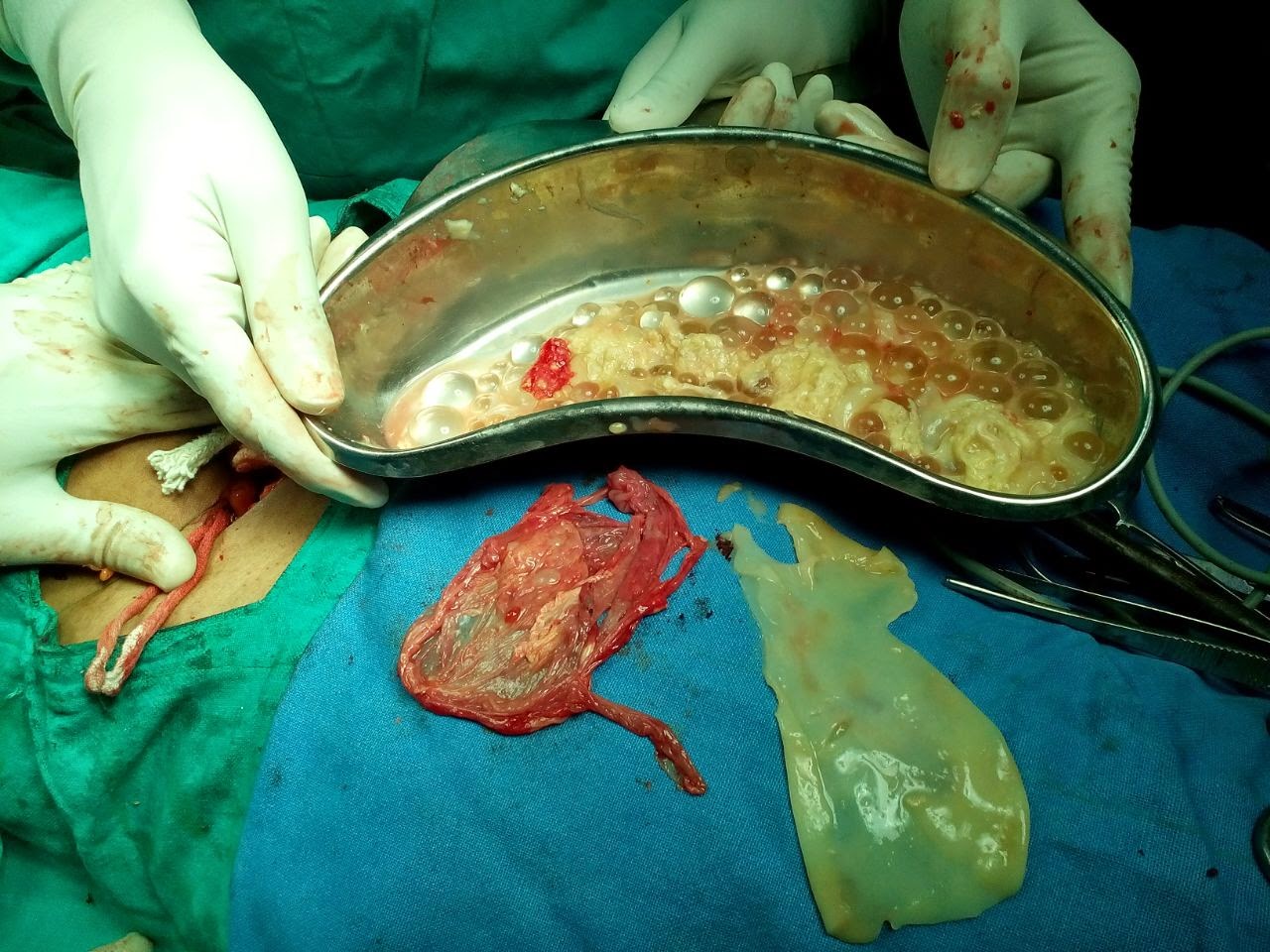
Fig 10- TVS pelvic scan shows large multiloculated adnexal mass with Honey Comb appearance.In this case mother cyst is full of multiple small daughter cysts. As both ovaries were seen separate and normal, the diagnosis of adnexal (likely broad ligament) pelvic hydatid cyst was made.
Fig 11- Post operative specimens of the above pelvic adnexal hydatid cyst case proved to be broad ligament hydatid cyst (one of the rare entity). Gross specimens showing ectocyst (red piece), endocyst (white piece) and kidney tray containing daughter cysts.
Fig 12- Hepatic sonogram shows a large rounded complex cystic mass in left lobe with Honey Comb appearance.
Fig 13-Hepatic sonogram shows a large complex cystic mass with multiple small daughter cysts see as mobile sediments, likely suggests detached daughter cysts.
Case 4 - CE 3 (WHO) type transitional stage hydatid cyst
.
Fig 14- Hepatic sonogram shows large complex cystic mass right lobe with detached endocyst layer appearing as echogenic curvilinear structure.
Fig 15- Hepatic sonogram of another case shows collapsed hydatid cyst with detached undulated endocyst.
Case 4- CE 4 type collapsed consolidating (solid appearing) inactive hydatid cyst.
Fig 16- Hepatic sonogram shows large rounded hyper echoic solid appearing mass with multiple hypoechoic curvilinear echos clustered within mass without daughter cysts. It is suggestive of consolidating inactive hydatid cyst.
Case 5 - Type 3 (morphological), CE 5 (WHO) type collapsed consolidating (solid appearing) inactive hydatid cyst.
Fig 17- Hepatic sonogram shows a well defined rounded mass with echogenic rim calcification casting distal shadowing. Here the lesion is inactive and infertile with calcifying nature.
Case 6 - Type 4 (morphological) Complicated hydatid cyst.
Fig 18- Hepatic Sonogram shows complex cystic mass left lobe liver full of internal echos and multiple bright foci with dirty down shadowing,suggestive of air. The case was earlier diagnosed as simple type hydatid cyst, which at present appears complicated by air and infection.
P.S.-This presentation is intended for medical professionals and imaging specialists for academic purposes. It needs discussion and further review of literature.
- My special thanks to Dr.Gaurav Bahety M.Ch. (Ped. Surgeon) and Dr.S P Sharma (Gen.Surgeon) for operative feedback, and Dr.Kartikeya Nathiya, Bhilwara, for article layout and crafting.
References
1] For more detail
study – ref. http://radiopaedia.org/articles/world-health-organization-2001-classification-of-hepatic-hydatid-cysts


.jpg)


.JPG)


