ENCYSTED /LOCULATED INFECTED PLEURAL EFFUSION [ EMPYEMA THORACIS]
Fig 1- Utrasound image of left side chest showing thick walled loculated pleural effusion with internal echos and septas - indicative of empyema .
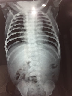
Fig 2- CXR PA of the same case showing homogeneous opacity left mid and lower zone with smooth upper margin . The opacity is obscuring cardiac and dome outline .
D/D - 1) Consolidation - point against - absent air bronchograms
2)- Pleural effusion - point against - no mass effect - this can be explained as it is loculated empyema
USG OF ENLARGED THYMUS
CXR: Shows upper mediastinal widening
USG scan(chest, left parmedian TS view) of the same pediatric case: Shows enlarged thymus as well defined soft tissue mass in the region of superior mediastinum retrosternal space.
USG scan(chest, left parmedian LS view) of the same pediatric case: Shows enlarged thymus as well defined soft tissue mass in the region of superior mediastinum retrosternal space, antero-superior to heart and great vessels.


