Tuesday, September 30, 2014

PELVIC ( BROAD LIGAMENT) HYDATID CYST- AN ULTRASOUND DIAGNOSIS

CASE DETAILS- A 20 yrs female with lower abdominal pain
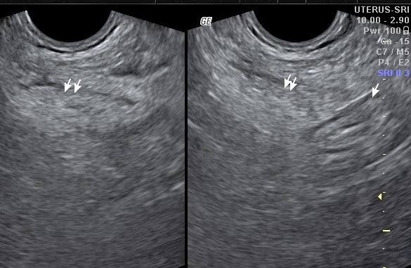
AT US -  A large well defined rounded complex multicystic  mass noted in pelvis separate from normally appearing uterus and ovaries. The mass was harbouring multiple small rounded cysts ranging10 to 20 mm in size , mostly similar,  like bunch of grapes, with echofree interior . The lesion was seen superior & slightly right to uterus and bladder. No internal echos or vascularity noted within mass . The mass was showing slight double layered mural appearance at periphery. No solid components seen . No free fluid was noted in POD. No calcific foci or any other internal complexity was appreciated . No any other similar lesion was seen elsewhere in abdomen. On the basis of US findings a diagnosis of benign complex pelvic cyst , possibly Hydatid cyst of right broad ligament [ primary pelvic peritoneal hydatid disease ],  was made. Which was confirmed at surgery.

Fig 1- TVS scan showing a large complex multicystic pelvic mass with multiple rounded daughter cysts in it, & the mass was separate from uterus & both ovaries


Fig 2 - TVS scan showing double layered peripheral wall of the multicystic mass

Fig 3 - TVS scans showing normally & separately appearing uterus & both ovaries

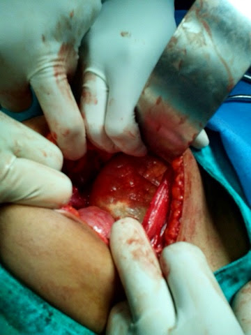
AT SURGERY- Rt broad ligament Hydatid cyst with thin filmy adhesion with right ovary, bladder wall, adjacent bowel loops & abdominal wall noted . The cyst was successfully operated and removed.
Fig 4 - Photograh showing per operative view of pelvic cyst 

Fig 5-  Post op gross specimen showing ectocyst layer ( red piece) , endocyst layer( creamy white piece) , & multiple small daughter cysts in kidney tray .

PS- 1) The case study is intended for medical professionals & imaging specialists for academic interests.
2) Special thanks to Dr S P sharma ( Surgeon, Bhilwara (raj, India) for operative feedback.

Friday, September 26, 2014

MULTIORGAN ULTRASOUND FINDINGS IN A CHILD WITH NONHODGKIN'S LYMPHOMA


CASE DETAILS-  A 6 yrs old boy presented with wt loss, pain abdomen, occasional vomiting & painless scrotal swelling. 
USG ABDOMEN- showed -
(1) Mild  hepatomegaly with multiple rounded hypoechoic solid nodular masses ranging 15 to 45 mm size involving both lobes. 
(2) A 6 cms long segment Jejunal loop hypoechoic concentric mural thickening ( 10 mm) in left upper abdomen with mild luminal dilatation ( classical aneurysmal dilatation with target sign ) . Rest bowel was normal.
(3) A large 4 cms sized rounded adjacent mesenteric nodal mass
(4) Slight omental thickening
(5) Mild ascites

Fig 1- Hepatic US scan shows multiple rounded hypoechoic nodular metastatic masses


Fig 2-  Left upper abdominal US scan shows hypoechoic concentric Jejunal loop thickening with aneurysmal dilatation. Adjacent rounded hypoechoic mesenteric nodal mass also seen.

SCROTAL USG - findings were -

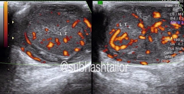
(1) Mildly bulky homogeneously hypoechoic both testes with mild hyperemia on doppler 
(2) Diffusely hypoechoic epididymis 
(3) Markedly thick inhomogenic hypoechoic & hyperemic extratesticular mass due to cord thickening, which is extending upto inguinal canal regions
(4) No hydrocele was present

Fig 3 - Scrotal US & color doppler scan showing homogeneously hypoechoic & hyperemic testis , & extratesticular masses due to cord & epididymal swelling.

Fig 4 - scrotal US scan showing hypoechoic testis & extratesticular masses due to cord and epididymal infiltration 

Fig 5- Quad US & color doppler images of both sided  supratesticular spermatic cords showing hypoechoic & hyperemic thickening of entire cord extending upto inguinal canal.

PS -1) The case study is intended for medical professionals and imaging specialists for academic purposes 
2) FNAC from hepatic nodule & testis showed NONHODGKIN'S LYMPHOMA 

Wednesday, September 10, 2014

ULTRASOUND DIAGNOSIS OF DUODENAL ULCER IN A PEDIATRIC CASE

CASE DETAILS : -
A 13 yrs old child with h/o chr pain upper abd , occasional vomiting , & had one episode of malena  , was referred for ultrasound . The case was evaluated with ultrasound with fasting status & allowed to sip some water to make upper GI window . The solid organs were normal . No small bowel dilatation was seen . The GEJ & stomach were also normal in mucosal pattern & appearance . The duodenal evaluation showed eccentric focal mural hypoechoic thickening ( 6 mm) in about15 to 20 mm circumferential area in its proximal part . There was a classical mucosal defect or focal depression noted in this region resembling an ulcer crater , harbouring specks of air foci in it ( fig 1). The lesion was constantly persistent on imaging  . Focal luminal & mural / mucosal distortion was also observed ( fig 1 ) . Multiple episodes of hyper duodeno-gastric regurgitation of fluid were also noted during real time scan . So on the basis of classical ultrasound findings a diagnosis of duodenal ulcer was made , which was subsequently confirmed on endoscopy ( fig 2 )
Fig 1- Epigastric midline slightly oblique US scan showing focal duodenal eccentric hypoechoic mural thickening encasing a classical depressive lesion s/o ulcer ( arrows)
Fig 2- Upper GI endoscopic images showing classical duodenal ulcer in frontal & profile views .
Fig 3- Collage showing US & Endoscopic images to highlight identical appearance of duodenal ulcer . 

PS - Endoscopic f/up : Dr S Bohra , Apollo Hospital , Ahmedabad .

Sunday, August 31, 2014

ULTRASOUND EVALUATION OF CA RECTUM

CASE HISTORY - A 40 yrs female with recurrent bleeding per rectum & lower abd pain .
AT USG -   On trans abd ultrasound focal iso to hypo & hyperechoic rounded lesions of variable size noted in liver with hypoechoic halo around ,s/o mets . Rest of abd scan was normal for abd viscera . To evaluate ano-colorectal region a non gyne TVS applied . Non gyne TVS application showed irregular hypoechoic mural thickening in rectum just above anorectal junction involving about 55 mm long segment with mural thickness ranging 10 to 14 mm distorting layer & mucosal pattern. The lesion was hyperemic on color Doppler. A tiny hypoechoic mural nodule also noted adjacently & separately   likely s/o satellite metastatic lesion . Multiple rounded hypoechoic perirectal lymphadenopathy also noted  . In view of above findings the diagnosis of Ca rectum was made with adjacent satellite mural lesion , perirectal metastatic lymphadenopathy  & hepatic metastasis .

Fig 1 - Hepatic scan showing mets

Fig 2- TVS scan with rectal evaluation showing perirectal nodes .

Fig 3- TVS scan showing irregular hypoechoic rectal wall thickening distorting gut signature  & adjacent tiny mural satellite lesion.


Fig 4- On color Doppler the rectal thickening showed hyperemia .
 
  This is how ultrasound completed the diagnosis & TVS helped in rectal evaluation in proper . 

PS - Histopathology revealed adeno ca rectum .

Sunday, August 24, 2014

ULTRASOUND IN NEONATAL HIRSCHSPRUNG DISEASE


CASE HISTORY 
A  20 days old neonate with distension of abdomen , at ultrasound showed  - slight narrowing of rectum ( dotted arrow )  & gross distension of sigmoid & descending colon ( arrow )  , & mild distension of entire colon. Rest of the abdomen was unremarkable  . On the basis of USG findings low (rectal) obstruction suspected with colonic distension , & possibility of Hirschsprung disease proposed .  On barium enema study classical findings of narrowing with coning ( zone of transition ) & distension of sigmoid noted , which supported further the diagnosis . The case underwent primary colostomy with rectal biopsy , which confirmed aganglionic nature Hirschsprung disease . 

Fig-1  suprapubic US scan shows rectal narrowing with coning ( dotted arrow ) , & distended sigmoid ( small arrow ) . UB seen empty . The image classically similar  to barium study .

Fig 2 - Quad US scan shows distended sigmoid , descending  & other parts of colon with rectal narrowing . 


Fig 3 - Lateral barium enema radiograph


Fig 4- AP barium enema radiograph 

Both fig 3 & 4 barium enema radiographs   
shows narrowing of rectum with coning / funnelling of zone of transition & distended sigmoid colon , classical for Hirschsprung  disease .  

Case 2-
Fig 5- Sono-Recto-sigmoidogram,showing rectal narrowing / stricture with sigmoid fullness-classically resembling with barium study consistent with possible diagnosis of "Hirschsprung disease " (story of a 30 days neonate with constipation & abdominal distension)

TAKE HOME- To diagnose Hirschsprung disease in neonatal period is quite feasible and rewarding. The liquid meconium in colon & recto-sigmoid region acts as a contrast ( echogenic liquid in bowel) during sonography & can delineate the rectal narrowing ( stricture as beak sign) with proximal sigmoid distension. In the late age the constipated & fecal matter loaded sigmoid obscures the stricture details due to fecal matter & gas shadowing .

PS - Barium enema radiographic images  & operative f/up courtesy : Dr Gaurav Bahety , M. Ch. pediatric surgeon , Bhilwara ( raj , India )


Thursday, July 10, 2014

OMENTAL INFARCT - ULTRSOUND EVALUATION

                                               POSSIBLE OMENTAL INFARCT

CLINICAL STORY - A 65 yrs old female  developped severe pain abdomen , and occasional vomiting.  Routine blood and urine lab tests were unremarkable . clinically there was a lumpish feel and local tenderness in right lower quadrant .

ULTRASOUND SCAN - Showed a slightly well defined  oval shaped hyperechoic mass of about 92x30x57mm size  in right lower abdomen , corresponding the site of tenderness [ Fig - 1 & 2] . The mass was lodged just deep to abdominal wall & did not reveal appreciable internal vascularity on doppler . No cystic or calcific foci seen . Underlying & adjacent small and large bowel loops were normal sonographically . Trace amount of ascites was present in right iliac fossa . Liver was sightly fatty . Right kidney & pelvic structure were also unremarkable . Appendix was not seen inflammed .  A probable diagnosis of inflammed fat , likey omental infarct was made , & CT scan was suggested .

CT SCAN -  Showed a well defined slightly heterogenic , predominently hypodese mass in right lower abdomen anterior to bowel loops with fat density areas [ Fig 3 &4 ] . No significant vascularity or ontrast uptake was seen . Rest of abdominal structures were unremarkable . A diagnosis of omental infarct was made on the basis of imaging findings .

 Fig - 1 , Sag. & transverse US image right lower quadrant shows an oval subtle 
                                      heterogenic hyperechoic mass just deep to anterior abdominal wall .

                                     Fig 2 - Appearance of the mass with high resolution linear probe

Fig 3


                                                                              Fig 4
Both fig 3 & fig 4 are CT images [both axial & sag reformation ] showing subtle heterogenic predominantly hypodense oval mass in right lower abdomen , anterior to bowel loops , with fat density areas , confirming the mass as - omental infarct
PS- the case study is intended for medical professionals & imaging specialists for academic purpose

Sunday, November 10, 2013

EPIPLOIC APPENDAGITIS - An ultrasound diagnosis

Epiploic  appendages are peritoneal pouches that arise from the serosal surface of the colon. These are composed of adipose tissue and attached to colon by vascular  stalk  , multiple in count [ approx 100 ] ,& have a length range of  about 0.5–5 cm. Those located near the sigmoid colon are the largest in size . Epiploic appendages are usually not found near the rectum. The appendages are arranged in two rows: one row medial to the tenia libera, and the other lateral to the tenia omentalis of the colon .


Epiploic appendagitis is an uncommon self limiting inflammatory or  ischaemic condition affecting the colonic  epiploic appendage. Appendagitis  may be primary[ spontaneous ] or secondary to adjacent pathology , with more affection or predilection for sigmoid colon , presumably due to larger appendices. The condition most commonly manifests in the 4th to 5th decades of life, predominantly in men & obese individuals . Torsion of epiploic appendage pedicle , with resultant vascular occlusion or venous occlusion that leads to ischemia[ ischemic necrosis], has been implicated as the cause of acute epiploic appendagitis.  Acute epiploic appendagitis is also seen to be associated with  hernia, and unaccustomed exercise. Mostly  it is a self limiting  disease  , and  very uncommonly it may result in adhesion, bowel obstruction, intussusception, intraperitoneal loose body, peritonitis, and/or focal abscess formation.


Clinically patients present with acute abdominal pain and guarding in local area at the site of involvement [ that is usually lower quadrant , more common in left iliac fossa] . On the basis of clinical manifestations alone, acute epiploic appendagitis is mis-diagnosed in most of the cases , and or  often its being mistaken for acute diverticulitis.  Unlike acute epiploic appendagitis, acute diverticulitis is more likely to cause evenly distributed lower abdominal pain and  associated with nausea, fever, and leukocytosis.  On the contrary most patients with acute epiploic appendagitis do not manifest  any change in their bowel habits, and only a few cases report constipation or diarrhea.

Differential diagnosis ;
1] acute omental infarction
2] diverticulitis
3]sclerosing mesenteritis or mesenteric panniculitis
4] primary tumor or metastasis that involves peritoneum and mesocolon.
Ultrasound Findings
The site of maximum tenderness[mostly left iliac fossa] is the region of interest for ultrasound evaluation . There is evidence of a round to oval echogenic  noncompressible  mass noted , with no internal vascularity on doppler, and surrounded by a subtle hypoechoic line . The lesion is  typically 2 - 5 cm in maximal diameter adjacent and anterior to colon , and just deep to abdominal wall . The mass exert slight compressive effect on colonic loop  , and usually not  associated with colonic mural thickening  , or local or free peritoneal fluid .
In my series of 7 patients  [ 6 M & 2F, age range 35 to 48 yrs] , six cases presented with pain left iliac fossa , & one case with pain in right iliac fossa.  All cases were showed classical ultrasound findings of epiploic appendagitis with no bowel thickening or ascites . In all cases no leucocytosis was observed in lab. tests. In one case with pain right iliac fossa , the d/d was with acute appendicitis , but appendix and ileo-cecal region was seen separately & normally on ultrasound  .

Fig 1- Shows a normal  descending colonic epiploic appendage : An oval elongated echogenic structure [ arrow] attached to DC loop, nicely depicted due to presence of ascites[FL]


Fig 2 - Epiploic appendagitis, case A : LIF scan shows a well defined oval echogenic mass [bet cursors] surrounded by hypoechoic halo , just deep to abd wall 

Fig 3 - LIF scan of same case A , mass with no internal vascularity on power  doppler

Fig 4 - CT scan sagittal reconstruction image of  case A, shows a well defined oval hypodense fat density lesion along descending colonic loop[arrow within circle]

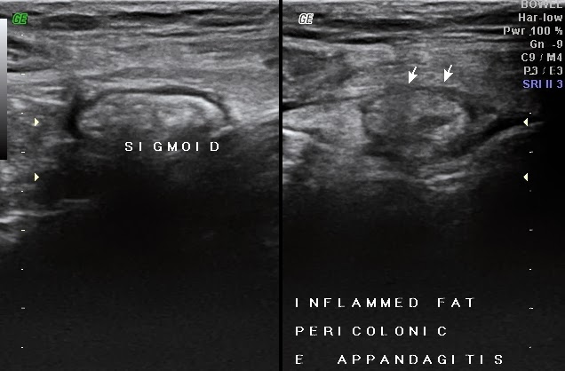
Fig 5 - Epiploic appendagitis, Case B, LIF scan shows a well defined  oval echogenic mass[ twin arrow] between sigmoid loop[single arrow] and abd wall

Fig 6 - Epiploic appendagitis, case C, LIF scan shows an oval echogenic mass with hypoechoic rim[arrows] adjacent to normally appearing sigmoid cooln


CT findings-
A fat-density ovoid lesion  adjacent to colon, size varying 1.5 - 3.5cm in diameter , with thin high-density rim , surrounding inflammatory fat stranding, and thickening of the adjacent peritoneum. A central hyperdense dot like area also appreciated representing the thrombosed vascular pedicle. As a chronic sequele an infarcted appendix epiploica  may calcify, and may detach to form an intraperitoneal loose body.

Treatment and prognosis

Epiploic appendagitis is a self limiting disease, and thus a correct diagnosis is important with imaging modalities to prevents unnecessary surgery. 
 Ref.